6AL2

Crystal structure of E. coli YidC at 2.8 A resolution


Experimental Data Snapshot

  • Method: X-RAY DIFFRACTION
  • Resolution: 2.80 Å
  • R-Value Free: 0.275 
  • R-Value Work: 0.218 
  • R-Value Observed: 0.223 

wwPDB Validation   3D Report Full Report


Ligand Structure Quality Assessment 


This is version 1.3 of the entry. See complete history


Literature

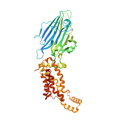
2.8-angstrom crystal structure of Escherichia coli YidC revealing all core regions, including flexible C2 loop.

Tanaka, Y.Izumioka, A.Abdul Hamid, A.Fujii, A.Haruyama, T.Furukawa, A.Tsukazaki, T.

(2018) Biochem Biophys Res Commun 505: 141-145

  • DOI: https://doi.org/10.1016/j.bbrc.2018.09.043
  • Primary Citation of Related Structures:  
    6AL2

  • PubMed Abstract: 

    YidC/Alb3/Oxa1 family proteins are involved in the insertion and assembly of membrane proteins. The core five transmembrane regions of YidC, which are conserved in the protein family, form a positively charged cavity open to the cytoplasmic side. The cavity plays an important role in membrane protein insertion. In all reported structural studies of YidC, the second cytoplasmic loop (C2 loop) was disordered, limiting the understanding of its role. Here, we determined the crystal structure of YidC including the C2 loop at 2.8 Å resolution with R/R free  = 21.8/27.5. This structure and subsequent molecular dynamics simulation indicated that the intrinsic flexible C2 loop covered the positively charged cavity. This crystal structure provides the coordinates of the complete core region including the C2 loop, which is valuable for further analyses of YidC.


  • Organizational Affiliation

    Graduate School of Science and Technology, Nara Institute of Science and Technology, Nara, 630-0192, Japan.


Macromolecules
Find similar proteins by:  (by identity cutoff)  |  3D Structure
Entity ID: 1
MoleculeChains Sequence LengthOrganismDetailsImage
Membrane protein insertase YidC
A, B
551Escherichia coliMutation(s): 0 
Gene Names: yidCECS88_4129
Membrane Entity: Yes 
UniProt
Find proteins for P25714 (Escherichia coli (strain K12))
Explore P25714 
Go to UniProtKB:  P25714
Entity Groups  
Sequence Clusters30% Identity50% Identity70% Identity90% Identity95% Identity100% Identity
UniProt GroupP25714
Sequence Annotations
Expand
  • Reference Sequence
Small Molecules
Ligands 1 Unique
IDChains Name / Formula / InChI Key2D Diagram3D Interactions
OLC
Query on OLC

Download Ideal Coordinates CCD File 
C [auth A](2R)-2,3-dihydroxypropyl (9Z)-octadec-9-enoate
C21 H40 O4
RZRNAYUHWVFMIP-GDCKJWNLSA-N
Experimental Data & Validation

Experimental Data

  • Method: X-RAY DIFFRACTION
  • Resolution: 2.80 Å
  • R-Value Free: 0.275 
  • R-Value Work: 0.218 
  • R-Value Observed: 0.223 
  • Space Group: P 1
Unit Cell:
Length ( Å )Angle ( ˚ )
a = 42.91α = 78.03
b = 76.19β = 82.61
c = 91.95γ = 78.05
Software Package:
Software NamePurpose
XDSdata reduction
XSCALEdata scaling
PHASERphasing
PHENIXrefinement
PDB_EXTRACTdata extraction

Structure Validation

View Full Validation Report


Ligand Structure Quality Assessment 


Entry History & Funding Information

Deposition Data


Funding OrganizationLocationGrant Number
Japan Society for the Promotion of ScienceJapanJP26119007
Japan Society for the Promotion of ScienceJapanJP26291023
Japan Society for the Promotion of ScienceJapanJP18H02405
Japan Society for the Promotion of ScienceJapanJP17H05669
Japan Society for the Promotion of ScienceJapanJP17K19528
Japan Society for the Promotion of ScienceJapanJP16K14713
Japan Society for the Promotion of ScienceJapanJP15H01537
Japan Society for the Promotion of ScienceJapanJP15K06972

Revision History  (Full details and data files)

  • Version 1.0: 2018-11-07
    Type: Initial release
  • Version 1.1: 2019-05-22
    Changes: Data collection, Structure summary
  • Version 1.2: 2020-03-04
    Changes: Data collection
  • Version 1.3: 2023-11-22
    Changes: Data collection, Database references, Refinement description