6AIS

Loop deletion mutant (deleting two residues)


Experimental Data Snapshot

  • Method: X-RAY DIFFRACTION
  • Resolution: 1.30 Å
  • R-Value Free: 0.203 
  • R-Value Work: 0.176 
  • R-Value Observed: 0.178 

wwPDB Validation   3D Report Full Report


This is version 1.2 of the entry. See complete history


Literature

Domain-Swapping Design by Polyproline Rod Insertion.

Shiga, S.Yamanaka, M.Fujiwara, W.Hirota, S.Goda, S.Makabe, K.

(2019) Chembiochem 20: 2454-2457

  • DOI: https://doi.org/10.1002/cbic.201900179
  • Primary Citation of Related Structures:  
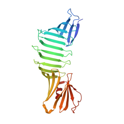
    6AIS, 6ICS, 6IDC, 6IEI, 6IYS

  • PubMed Abstract: 

    During domain swapping, proteins mutually interconvert structural elements to form a di-/oligomer. Engineering this process by design is important for creating a higher order protein assembly with minimal modification. Herein, a simple design strategy is shown for domain-swapping formation by loop deletion and insertion of a polyproline rod. Crystal structures revealed the formation of the domain-swapped dimers and polyproline portion formed a polyproline II (PPII) structure. Small-angle X-ray scattering demonstrated that an extended orientation of domain-swapped dimer was retained in solution. It is found that a multiple of three of inserting proline residue is favored for domain swapping because of the helical nature of PPII. The rigid nature of the polyproline rod enables precise control of the interdomain distance and orientation.


  • Organizational Affiliation

    Graduate School of Science and Engineering, Yamagata University, Jyonan 4-3-16, Yonezawa, Yamagata, 992-8510, Japan.


Macromolecules
Find similar proteins by:  (by identity cutoff)  |  3D Structure
Entity ID: 1
MoleculeChains Sequence LengthOrganismDetailsImage
Outer surface protein AA [auth O]249Borreliella burgdorferiMutation(s): 0 
Entity Groups  
Sequence Clusters30% Identity50% Identity70% Identity90% Identity95% Identity100% Identity
Sequence Annotations
Expand
  • Reference Sequence
Experimental Data & Validation

Experimental Data

  • Method: X-RAY DIFFRACTION
  • Resolution: 1.30 Å
  • R-Value Free: 0.203 
  • R-Value Work: 0.176 
  • R-Value Observed: 0.178 
  • Space Group: P 1 21 1
Unit Cell:
Length ( Å )Angle ( ˚ )
a = 33.003α = 90
b = 54.876β = 100.54
c = 66.19γ = 90
Software Package:
Software NamePurpose
PHENIXrefinement
HKL-2000data reduction
HKL-2000data scaling
Cootmodel building
MOLREPphasing

Structure Validation

View Full Validation Report


Entry History 

Deposition Data

Revision History  (Full details and data files)

  • Version 1.0: 2019-07-03
    Type: Initial release
  • Version 1.1: 2019-10-16
    Changes: Data collection, Database references
  • Version 1.2: 2023-11-22
    Changes: Data collection, Database references, Refinement description