6ABI

The apo-structure of D-lactate dehydrogenase from Fusobacterium nucleatum


Experimental Data Snapshot

  • Method: X-RAY DIFFRACTION
  • Resolution: 2.10 Å
  • R-Value Free: 0.217 
  • R-Value Work: 0.186 
  • R-Value Observed: 0.188 

wwPDB Validation   3D Report Full Report


This is version 1.3 of the entry. See complete history

Re-refinement Note

This entry reflects an alternative modeling of the original data in: 3WWY


Literature

Structural Basis of Sequential Allosteric Transitions in Tetrameric d-Lactate Dehydrogenases from Three Gram-Negative Bacteria

Furukawa, N.Miyanaga, A.Nakajima, M.Taguchi, H.

(2018) Biochemistry 57: 5388-5406

  • DOI: https://doi.org/10.1021/acs.biochem.8b00557
  • Primary Citation of Related Structures:  
    5Z1Z, 5Z20, 5Z21, 6ABI, 6ABJ

  • PubMed Abstract: 

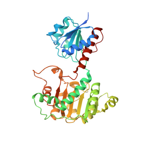
    d-Lactate dehydrogenases (d-LDHs) from Fusobacterium nucleatum (FnLDH) and Escherichia coli (EcLDH) exhibit positive cooperativity in substrate binding, and the Pseudomonas aeruginosa enzyme (PaLDH) shows negatively cooperative substrate binding. The apo and ternary complex structures of FnLDH and PaLDH have been determined together with the apo-EcLDH structure. The three enzymes consistently form homotetrameric structures with three symmetric axes, the P-, Q-, and R-axes, unlike Lactobacillus d-LDHs, P-axis-related dimeric enzymes, although apo-FnLDH and EcLDH form asymmetric and distorted quaternary structures. The tetrameric structure allows apo-FnLDH and EcLDH to form wide intersubunit contact surfaces between the opened catalytic domains of the two Q-axis-related subunits in coordination with their asymmetric and distorted quaternary structures. These contact surfaces comprise intersubunit hydrogen bonds and hydrophobic interactions and likely prevent the domain closure motion during initial substrate binding. In contrast, apo-PaLDH possesses a highly symmetrical quaternary structure and partially closed catalytic domains that are favorable for initial substrate binding and forms virtually no intersubunit contact surface between the catalytic domains, which present their negatively charged surfaces to each other at the subunit interface. Complex FnLDH and PaLDH possess highly symmetrical quaternary structures with closed forms of the catalytic domains, which are separate from each other at the subunit interface. Structure-based mutations successfully converted the three enzymes to their dimeric forms, which exhibited no significant cooperativity in substrate binding. These observations indicate that the three enzymes undergo typical sequential allosteric transitions to exhibit their distinctive allosteric functions through the tetrameric structures.


  • Organizational Affiliation

    Department of Applied Biological Science, Faculty of Science and Technology , Tokyo University of Science , 2641 Yamazaki , Noda , Chiba 278-8510 , Japan.


Macromolecules
Find similar proteins by:  (by identity cutoff)  |  3D Structure
Entity ID: 1
MoleculeChains Sequence LengthOrganismDetailsImage
D-lactate dehydrogenase
A, B
358Fusobacterium nucleatum subsp. nucleatum ATCC 25586Mutation(s): 0 
Gene Names: FN0511
EC: 1.1.1.28
UniProt
Find proteins for Q8RG11 (Fusobacterium nucleatum subsp. nucleatum (strain ATCC 25586 / DSM 15643 / BCRC 10681 / CIP 101130 / JCM 8532 / KCTC 2640 / LMG 13131 / VPI 4355))
Explore Q8RG11 
Go to UniProtKB:  Q8RG11
Entity Groups  
Sequence Clusters30% Identity50% Identity70% Identity90% Identity95% Identity100% Identity
UniProt GroupQ8RG11
Sequence Annotations
Expand
  • Reference Sequence
Small Molecules
Ligands 2 Unique
IDChains Name / Formula / InChI Key2D Diagram3D Interactions
SO4
Query on SO4

Download Ideal Coordinates CCD File 
C [auth A]
D [auth A]
E [auth A]
F [auth A]
H [auth B]
C [auth A],
D [auth A],
E [auth A],
F [auth A],
H [auth B],
I [auth B],
J [auth B],
K [auth B]
SULFATE ION
O4 S
QAOWNCQODCNURD-UHFFFAOYSA-L
GOL
Query on GOL

Download Ideal Coordinates CCD File 
G [auth A]
L [auth B]
M [auth B]
N [auth B]
O [auth B]
G [auth A],
L [auth B],
M [auth B],
N [auth B],
O [auth B],
P [auth B],
Q [auth B],
R [auth B]
GLYCEROL
C3 H8 O3
PEDCQBHIVMGVHV-UHFFFAOYSA-N
Experimental Data & Validation

Experimental Data

  • Method: X-RAY DIFFRACTION
  • Resolution: 2.10 Å
  • R-Value Free: 0.217 
  • R-Value Work: 0.186 
  • R-Value Observed: 0.188 
  • Space Group: P 43 21 2
Unit Cell:
Length ( Å )Angle ( ˚ )
a = 116.29α = 90
b = 116.29β = 90
c = 234.4γ = 90
Software Package:
Software NamePurpose
REFMACrefinement
ADSCdata collection
SCALAdata scaling
ARP/wARPmodel building
MOLREPphasing
iMOSFLMdata reduction

Structure Validation

View Full Validation Report


Entry History 

Deposition Data

Revision History  (Full details and data files)

  • Version 1.0: 2018-09-19
    Type: Initial release
  • Version 1.1: 2018-09-26
    Changes: Data collection, Database references
  • Version 1.2: 2022-04-27
    Changes: Database references
  • Version 1.3: 2023-11-22
    Changes: Data collection, Refinement description