Structural basis for specific recognition of core fucosylation in N-glycans by Pholiota squarrosa lectin (PhoSL).
Yamasaki, K., Kubota, T., Yamasaki, T., Nagashima, I., Shimizu, H., Terada, R.I., Nishigami, H., Kang, J., Tateno, M., Tateno, H.(2019) Glycobiology 29: 576-587
- PubMed: 30913288
- DOI: https://doi.org/10.1093/glycob/cwz025
- Primary Citation of Related Structures:
6A86, 6A87 - PubMed Abstract:
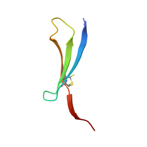
Fucosylation of the N-glycan core via the α1-6 linkage (core fucosylation) is detected in specific types of cancers and related diseases, and thereby serves for a relevant biomarker. The lectin from a mushroom Pholiota squarrosa (PhoSL) shows a clear specificity to core fucosylation, without recognizing those with other types of fucosylation, such as the H type via the α1-2 linkage or the Lewis type via the α1-3 or α1-4 linkage. Here we determined the crystal structure of the PhoSL trimer in complex with a disaccharide fucose(α1-6)N-acetylglucosamine (GlcNAc). In the three sugar-binding pockets of PhoSL, extensive hydrophobic and hydrogen-bonding contacts were formed with the fucose moiety. In contrast, the GlcNAc moiety showed only a few hydrophobic and hydrogen-bonding contacts. To elucidate the mechanism for the specificity, we performed molecular dynamics simulations on this disaccharide and a trisaccharide fucose(α1-6)[GlcNAc(β1-4)]GlcNAc in complex with PhoSL. It was observed that the GlcNAc corresponding to the outer one of the N-glycan core entered the sugar-binding pocket with the N-acetyl group placed stably at the bottom, forming extensive hydrophobic and hydrogen-bonding interactions. In addition, these glycans adopted unstressed favorable conformations when bound to PhoSL. In contrast, H- and Lewis-types of fucosylated trisaccharides adopting favorable conformations caused inevitable steric hindrance with the steep edge of the binding pocket, when docked with PhoSL. Therefore, the specificity to core fucosylation of PhoSL was achieved by a combination of these preferential and exclusive mechanisms.
Organizational Affiliation:
Biomedical Research Institute, National Institute of Advanced Industrial Science and Technology (AIST), 1-1-1 Higashi, Tsukuba, Japan.