Structural basis of the cystein protease inhibitor Clonorchis sinensis Stefin-1.
Park, S.Y., Jeong, M.S., Park, S.A., Ha, S.C., Na, B.K., Jang, S.B.(2018) Biochem Biophys Res Commun 498: 9-17
- PubMed: 29499196
- DOI: https://doi.org/10.1016/j.bbrc.2018.02.196
- Primary Citation of Related Structures:
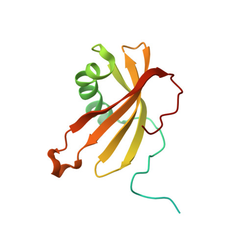
5ZC1 - PubMed Abstract:
Cystein protease plays a critical role as a virulence factor in the development and progression of various diseases. Cystatin is a superfamily of cysteine protease inhibitors that participates in various physiological and pathological processes. The cysteine protease inhibitor CsStein-1 isolated from Clonorchis sinensis belongs to the type 1 stefin of cystatins. This inhibitor regulates the activity and processing of CsCF (Cathepsin F of Clonorchis sienesis), which plays an important role in parasite nutrition and host-parasite interaction. CsStefin-1 has also been proposed as a host immune modulator and a participant in the mechanism associated with anti-inflammatory ability. Here, we report the first crystal structure of CsStefin-1 determined by the multi-wavelength anomalous diffraction (MAD) method to 2.3 Å. There are six molecules of CsStefin-1 per asymmetric unit, with a solvent content of 36.5%. The structure of CsStefin-1 is composed of twisted four-stranded antiparallel β-sheets, a central α-helix, and a short α-helix. We also demonstrate that CsStefin-1 binds to CsCF-8 cysteine protease and inhibits its activity. In addition, a molecular docking model of CsStefin-1 and CsCF-8 was developed using homology modeling based on their structures. The structural information regarding CsStefin-1 and molecular insight into its interaction with CsCF-8 are important to understanding their biological function and to design of inhibitors that modulate cysteine protease activity.
Organizational Affiliation:
Department of Molecular Biology, College of Natural Sciences, Pusan National University, Jangjeon-dong, Geumjeong-gu, Busan, 46241, Republic of Korea.