5YUA

Crystal structure of voltage-gated sodium channel NavAb in high-pH condition


Experimental Data Snapshot

  • Method: X-RAY DIFFRACTION
  • Resolution: 2.80 Å
  • R-Value Free: 0.262 
  • R-Value Work: 0.230 
  • R-Value Observed: 0.232 

wwPDB Validation   3D Report Full Report


Ligand Structure Quality Assessment 


This is version 1.2 of the entry. See complete history


Literature

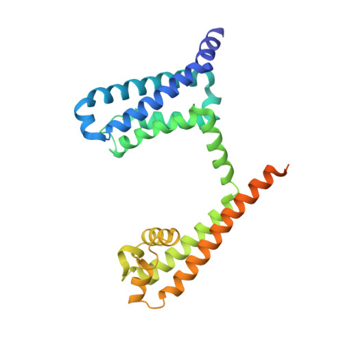
Structural insight on the voltage dependence of prokaryotic voltage gated sodium channel NavAb.

Irie, K.Haga, Y.Shimomura, T.Fujiyoshi, Y.

(2017) FEBS Lett 

  • DOI: https://doi.org/10.1002/1873-3468.12955
  • Primary Citation of Related Structures:  
    5YUA, 5YUB, 5YUC

  • PubMed Abstract: 

    Voltage-gated sodium channels are crucial for electro-signalling in living systems. Analysis of the molecular mechanism requires both fine electrophysiological evaluation and high-resolution channel structures. Here, we optimized a dual expression system of NavAb, which is a well-established standard of prokaryotic voltage-gated sodium channels, for E. coli and insect cells using a single plasmid vector to analyse high-resolution protein structures and measure large ionic currents. Using this expression system, we evaluated the voltage dependence and determined the crystal structures of NavAb wild-type and two mutants, E32Q and N49K, whose voltage dependence were positively shifted and essential interactions were lost in voltage sensor domain. The structural and functional comparison elucidated the molecular mechanisms of the voltage dependence of prokaryotic voltage-gated sodium channels.


  • Organizational Affiliation

    Cellular and Structural Physiology Institute (CeSPI), Nagoya University, Japan.


Macromolecules
Find similar proteins by:  (by identity cutoff)  |  3D Structure
Entity ID: 1
MoleculeChains Sequence LengthOrganismDetailsImage
Ion transport protein271Aliarcobacter butzleriMutation(s): 0 
Gene Names: SAMEA4475700_01771
Membrane Entity: Yes 
UniProt
Find proteins for A8EVM5 (Aliarcobacter butzleri (strain RM4018))
Explore A8EVM5 
Go to UniProtKB:  A8EVM5
Entity Groups  
Sequence Clusters30% Identity50% Identity70% Identity90% Identity95% Identity100% Identity
UniProt GroupA8EVM5
Sequence Annotations
Expand
  • Reference Sequence
Small Molecules
Ligands 4 Unique
IDChains Name / Formula / InChI Key2D Diagram3D Interactions
PX4
Query on PX4

Download Ideal Coordinates CCD File 
F [auth A]
G [auth A]
H [auth A]
I [auth A]
J [auth A]
F [auth A],
G [auth A],
H [auth A],
I [auth A],
J [auth A],
K [auth A]
1,2-DIMYRISTOYL-SN-GLYCERO-3-PHOSPHOCHOLINE
C36 H73 N O8 P
CITHEXJVPOWHKC-UUWRZZSWSA-O
1N7
Query on 1N7

Download Ideal Coordinates CCD File 
E [auth A]CHAPSO
C32 H59 N2 O8 S
GUQQBLRVXOUDTN-XOHPMCGNSA-O
LMT
Query on LMT

Download Ideal Coordinates CCD File 
C [auth A],
D [auth A]
DODECYL-BETA-D-MALTOSIDE
C24 H46 O11
NLEBIOOXCVAHBD-QKMCSOCLSA-N
CA
Query on CA

Download Ideal Coordinates CCD File 
B [auth A]CALCIUM ION
Ca
BHPQYMZQTOCNFJ-UHFFFAOYSA-N
Experimental Data & Validation

Experimental Data

  • Method: X-RAY DIFFRACTION
  • Resolution: 2.80 Å
  • R-Value Free: 0.262 
  • R-Value Work: 0.230 
  • R-Value Observed: 0.232 
  • Space Group: I 4 2 2
Unit Cell:
Length ( Å )Angle ( ˚ )
a = 126.45α = 90
b = 126.45β = 90
c = 201.658γ = 90
Software Package:
Software NamePurpose
PHENIXrefinement
HKL-2000data reduction
SCALEPACKdata scaling
PHASERphasing

Structure Validation

View Full Validation Report


Ligand Structure Quality Assessment 


Entry History & Funding Information

Deposition Data


Funding OrganizationLocationGrant Number
Toyoaki Scholarship FoundationJapan--

Revision History  (Full details and data files)

  • Version 1.0: 2017-12-27
    Type: Initial release
  • Version 1.1: 2018-01-17
    Changes: Database references
  • Version 1.2: 2023-11-22
    Changes: Data collection, Database references, Refinement description