5YNE

Crystal structure of Pullulanase from Klebsiella pneumoniae complex at 10 mM alpha-cyclodextrin


Experimental Data Snapshot

  • Method: X-RAY DIFFRACTION
  • Resolution: 2.20 Å
  • R-Value Free: 0.242 
  • R-Value Work: 0.181 
  • R-Value Observed: 0.184 

wwPDB Validation   3D Report Full Report


This is version 2.1 of the entry. See complete history


Literature

Elucidation of the mechanism of interaction between Klebsiella pneumoniae pullulanase and cyclodextrin

Saka, N.Iwamoto, H.Malle, D.Takahashi, N.Mizutani, K.Mikami, B.

(2018) Acta Crystallogr D Struct Biol 74: 1115-1123

  • DOI: https://doi.org/10.1107/S2059798318014523
  • Primary Citation of Related Structures:  
    5YN2, 5YN7, 5YNA, 5YNC, 5YND, 5YNE, 5YNH

  • PubMed Abstract: 

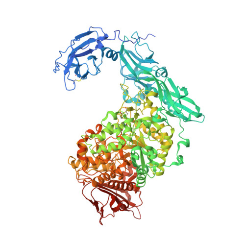
    Crystal structures of Klebsiella pneumoniae pullulanase (KPP) in complex with α-cyclodextrin (α-CD), β-cyclodextrin (β-CD) and γ-cyclodextrin (γ-CD) were refined at around 1.98-2.59 Å resolution from data collected at SPring-8. In the structures of the complexes obtained with 1 mM α-CD or γ-CD, one molecule of CD was found at carbohydrate-binding module 41 only (CBM41). In the structures of the complexes obtained with 1 mM β-CD or with 10 mM α-CD or γ-CD, two molecules of CD were found at CBM41 and in the active-site cleft, where the hydrophobic residue of Phe746 occupies the inside cavity of the CD rings. In contrast to α-CD and γ-CD, one β-CD molecule was found at the active site only in the presence of 0.1 mM β-CD. These results were coincident with the solution experiments, which showed that β-CD inhibits this enzyme more than a thousand times more potently than α-CD and γ-CD. The strong inhibition of β-CD is caused by the optimized interaction between β-CD and the side chain of Phe746. The increased K i values of the F746A mutant for β-CD supported the importance of Phe746 in the strong interaction of pullulanase with β-CD.


  • Organizational Affiliation

    Laboratory of Applied Structural Biology, Division of Applied Life Sciences, Graduate School of Agriculture, Kyoto University, Gokasyo, Uji, Kyoto 611-0011, Japan.


Macromolecules
Find similar proteins by:  (by identity cutoff)  |  3D Structure
Entity ID: 1
MoleculeChains Sequence LengthOrganismDetailsImage
PulA protein1,102Klebsiella pneumoniaeMutation(s): 0 
UniProt
Find proteins for W9BQ28 (Klebsiella pneumoniae)
Explore W9BQ28 
Go to UniProtKB:  W9BQ28
Entity Groups  
Sequence Clusters30% Identity50% Identity70% Identity90% Identity95% Identity100% Identity
UniProt GroupW9BQ28
Sequence Annotations
Expand
  • Reference Sequence
Oligosaccharides

Help

Entity ID: 2
MoleculeChains Length2D Diagram Glycosylation3D Interactions
Cyclohexakis-(1-4)-(alpha-D-glucopyranose)
B, C
6N/A
Glycosylation Resources
GlyTouCan:  G22307HL
GlyCosmos:  G22307HL
Small Molecules
Ligands 3 Unique
IDChains Name / Formula / InChI Key2D Diagram3D Interactions
PEG
Query on PEG

Download Ideal Coordinates CCD File 
O [auth A]DI(HYDROXYETHYL)ETHER
C4 H10 O3
MTHSVFCYNBDYFN-UHFFFAOYSA-N
GOL
Query on GOL

Download Ideal Coordinates CCD File 
I [auth A]
J [auth A]
K [auth A]
L [auth A]
M [auth A]
I [auth A],
J [auth A],
K [auth A],
L [auth A],
M [auth A],
N [auth A]
GLYCEROL
C3 H8 O3
PEDCQBHIVMGVHV-UHFFFAOYSA-N
CA
Query on CA

Download Ideal Coordinates CCD File 
D [auth A],
E [auth A],
F [auth A],
G [auth A],
H [auth A]
CALCIUM ION
Ca
BHPQYMZQTOCNFJ-UHFFFAOYSA-N
Biologically Interesting Molecules (External Reference) 1 Unique
Experimental Data & Validation

Experimental Data

  • Method: X-RAY DIFFRACTION
  • Resolution: 2.20 Å
  • R-Value Free: 0.242 
  • R-Value Work: 0.181 
  • R-Value Observed: 0.184 
  • Space Group: P 43 21 2
Unit Cell:
Length ( Å )Angle ( ˚ )
a = 89.21α = 90
b = 89.21β = 90
c = 299.647γ = 90
Software Package:
Software NamePurpose
HKL-2000data collection
MOLREPphasing
PHENIXrefinement
DENZOdata reduction
SCALEPACKdata scaling

Structure Validation

View Full Validation Report


Entry History & Funding Information

Deposition Data


Funding OrganizationLocationGrant Number
Japan Society for the Promotion of ScienceJapan16H04909

Revision History  (Full details and data files)

  • Version 1.0: 2018-10-24
    Type: Initial release
  • Version 1.1: 2018-11-14
    Changes: Data collection, Database references
  • Version 2.0: 2020-07-29
    Type: Remediation
    Reason: Carbohydrate remediation
    Changes: Atomic model, Data collection, Derived calculations, Structure summary
  • Version 2.1: 2023-11-22
    Changes: Data collection, Database references, Derived calculations, Refinement description, Structure summary