X-ray structure of Clostridium perfringens sortase B cysteine transpeptidase
Tamai, E., Sekiya, H., Maki, J., Nariya, H., Yoshida, H., Kamitori, S.(2017) Biochem Biophys Res Commun 493: 1267-1272
- PubMed: 28962862
- DOI: https://doi.org/10.1016/j.bbrc.2017.09.144
- Primary Citation of Related Structures:
5B23, 5YFK - PubMed Abstract:
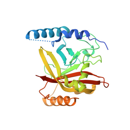
The pathogenesis and infectivity of Gram-positive bacteria are mediated by many surface proteins that are covalently attached to peptidoglycans of the cell wall. The covalent attachment of these proteins is catalyzed by sortases (Srts), a family of cysteine transpeptidases, which are classified into six classes, A - F, based on their amino acid sequences and biological roles. Clostridium perfringens, one of the pathogenic clostridial species, has a class B sortase (CpSrtB) with 249 amino acid residues. X-ray structures of CpSrtB and its inactive mutant form were determined at 2.2 Å and 1.8 Å resolutions, respectively. CpSrtB adopts a typical sortase-protein fold, and has a unique substrate-binding groove formed by three β-strands and two helices creating the sidewalls of the groove. The position of the catalytic Cys232 of CpSrtB is significantly different from those commonly found in Srts structures. The modeling study of the CpSrtB/peptide complex suggested that the position of Cys232 found in CpSrtB is preferable for the catalytic reaction to occur. Structural comparison with other class B sortases demonstrated that the catalytic site likely converts between two forms. The movement of Cys232 between the two forms may help His136 deprotonate Cys232 to be activated as a thiolate, which may the catalytic Cys-activated mechanism for Srts.
Organizational Affiliation:
Department of Infectious Disease, College of Pharmaceutical Science, Matsuyama University, 4-2 Bunkyo-cho, Matsuyama, Ehime 790-8578, Japan; Life Science Research Center and Faculty of Medicine, Kagawa University, 1750-1 Ikenobe, Miki-cho, Kita-gun, Kagawa 761-0793, Japan.