


Crystal structure of Poecilia reticulata adenylate kinase
Bae, E., Kim, J., Moon, S.To be published.
Experimental Data Snapshot
Entity ID: 1 | |||||
|---|---|---|---|---|---|
| Molecule | Chains | Sequence Length | Organism | Details | Image |
| Adenylayte kinase | 193 | Poecilia reticulata | Mutation(s): 0 | 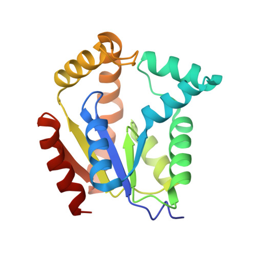 | |
UniProt | |||||
Find proteins for A0A452CSM2 (Poecilia reticulata) Explore A0A452CSM2 Go to UniProtKB: A0A452CSM2 | |||||
Entity Groups | |||||
| Sequence Clusters | 30% Identity50% Identity70% Identity90% Identity95% Identity100% Identity | ||||
| UniProt Group | A0A452CSM2 | ||||
Sequence AnnotationsExpand | |||||
| |||||
| Ligands 2 Unique | |||||
|---|---|---|---|---|---|
| ID | Chains | Name / Formula / InChI Key | 2D Diagram | 3D Interactions | |
| AP5 Query on AP5 | C [auth A], E [auth B] | BIS(ADENOSINE)-5'-PENTAPHOSPHATE C20 H29 N10 O22 P5 OIMACDRJUANHTJ-XPWFQUROSA-N |  | ||
| MG Query on MG | D [auth A], F [auth B] | MAGNESIUM ION Mg JLVVSXFLKOJNIY-UHFFFAOYSA-N |  | ||
| Length ( Å ) | Angle ( ˚ ) |
|---|---|
| a = 40.79 | α = 90 |
| b = 81.505 | β = 90 |
| c = 120.123 | γ = 90 |
| Software Name | Purpose |
|---|---|
| PHENIX | refinement |
| HKL-2000 | data reduction |
| HKL-2000 | data scaling |
| PHENIX | phasing |
| Funding Organization | Location | Grant Number |
|---|---|---|
| Rural Development Administration | Korea, Republic Of | PJ01111201 |
| National Research Foundation of Korea | Korea, Republic Of | NRF-2016R1D1A1A09916821 |
RCSB PDB (citation) is hosted by
RCSB PDB is a member of the

