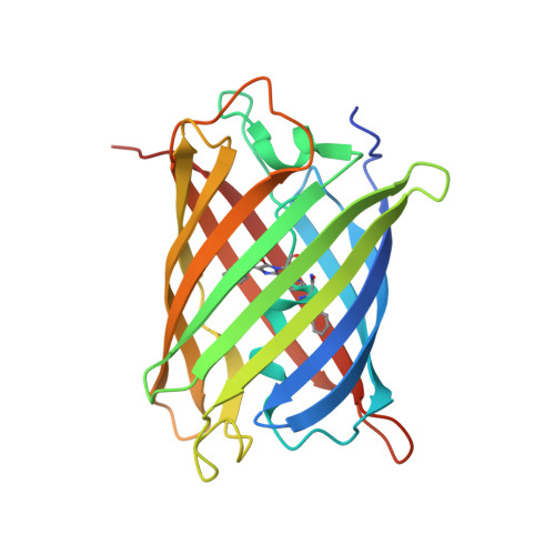
Disruption of the hydrogen bonding network determines the pH-induced non-fluorescent state of the fluorescent protein ZsYellow by protonation of Glu221.
Bae, J.E., Kim, I.J., Nam, K.H.(2017) Biochem Biophys Res Commun 493: 562-567
- PubMed: 28867188
- DOI: https://doi.org/10.1016/j.bbrc.2017.08.152
- Primary Citation of Related Structures:
5Y8Q, 5Y8R - PubMed Abstract:
Many fluorescent proteins (FPs) exhibit fluorescence quenching at a low pH. This pH-induced non-fluorescent state of an FP serves as a useful indicator of the cellular pH. ZsYellow is widely used as an optical marker in molecular biology, but its pH-induced non-fluorescent state has not been characterized. Here, we report the pH-dependent spectral properties of ZsYellow, which exhibited the pH-induced non-fluorescence state at a pH below 4.0. We determined the crystal structures of ZsYellow at pH 3.5 (non-fluorescence state) and 8.0 (fluorescence state), which revealed the cis-configuration of the chromophore without pH-induced isomerization. In the non-fluorescence state, Arg95, which is involved in stabilization of the exited state of the chromophore, was found to more loosely interact with the carbonyl oxygen atom of the chromophore when compared to the interaction at pH 8.0. In the fluorescence state, Glu221, which is involved in the hydrogen bonding network around the chromophore, stably interacted with Gln42 and His202. By contrast, in the non-fluorescence state, the protonated conserved Glu221 residue exhibited a large conformational change and was separated from His202 by 5.46 Å, resulting in breakdown of the hydrogen bond network. Our results provide insight into the critical role of the conserved Glu221 residue for generating the pH-induced non-fluorescent state.
Organizational Affiliation:
Pohang Accelerator Laboratory, Pohang University of Science and Technology, Pohang, 35398, Republic of Korea; School of Life Sciences, Kyungpook National University, Daegu, 41566, Republic of Korea.