5XTF

Crystal structure of the cis-dihydrodiol naphthalene dehydrogenase NahB from Pseudomonas sp. MC1


Experimental Data Snapshot

  • Method: X-RAY DIFFRACTION
  • Resolution: 2.10 Å
  • R-Value Free: 0.244 
  • R-Value Work: 0.204 
  • R-Value Observed: 0.206 

wwPDB Validation   3D Report Full Report


This is version 1.2 of the entry. See complete history


Literature

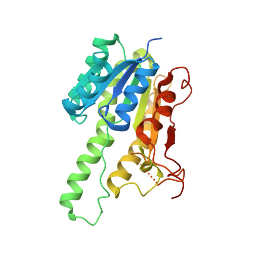
Crystal structure of cis-dihydrodiol naphthalene dehydrogenase (NahB) from Pseudomonas sp. MC1: Insights into the early binding process of the substrate

Park, A.K.Kim, H.Kim, I.S.Roh, S.J.Shin, S.C.Lee, J.H.Park, H.Kim, H.W.

(2017) Biochem Biophys Res Commun 491: 403-408

  • DOI: https://doi.org/10.1016/j.bbrc.2017.07.089
  • Primary Citation of Related Structures:  
    5XTF, 5XTG

  • PubMed Abstract: 

    The bacterial strain Pseudomonas sp. MC1 harbors an 81-kb metabolic plasmid, which encodes enzymes involved in the conversion of naphthalene to salicylate. Of these, the enzyme NahB (cis-dihydrodiol naphthalene dehydrogenase), which catalyzes the second reaction of this pathway, binds to various substrates such as cis-1,2-dihydro-1,2-dihydroxy-naphthalene (1,2-DDN), cis-2,3-dihydro-2,3-dihydroxybiphenyl (2,3-DDB), and 3,4-dihydro-3,4-dihydroxy-2,2',5,5'-tetrachlorobiphenyl (3,4-DD-2,2',5-5-TCB). However, the mechanism underlying its broad substrate specificity is unclear owing to the lack of structural information. Here, we determined the first crystal structures of NahB in the absence and presence of NAD + and 2,3-dihydroxybiphenyl (2,3-DB). Structure analysis suggests that the flexible substrate-binding loop allows NahB to accommodate diverse substrates. Furthermore, we defined the initial steps of substrate recognition and identified the early substrate-binding site in the substrate recognition process through the complex structure with ligands.


  • Organizational Affiliation

    Unit of Polar Genomics, Korea Polar Research Institute, Incheon 21990, South Korea.


Macromolecules
Find similar proteins by:  (by identity cutoff)  |  3D Structure
Entity ID: 1
MoleculeChains Sequence LengthOrganismDetailsImage
2,3-dihydroxy-2,3-dihydrophenylpropionate dehydrogenase
A, B
278Pseudomonas sp. MC1Mutation(s): 0 
Gene Names: nahBpYIC1_37
UniProt
Find proteins for G9G7I7 (Pseudomonas sp. MC1)
Explore G9G7I7 
Go to UniProtKB:  G9G7I7
Entity Groups  
Sequence Clusters30% Identity50% Identity70% Identity90% Identity95% Identity100% Identity
UniProt GroupG9G7I7
Sequence Annotations
Expand
  • Reference Sequence
Experimental Data & Validation

Experimental Data

  • Method: X-RAY DIFFRACTION
  • Resolution: 2.10 Å
  • R-Value Free: 0.244 
  • R-Value Work: 0.204 
  • R-Value Observed: 0.206 
  • Space Group: P 2 21 21
Unit Cell:
Length ( Å )Angle ( ˚ )
a = 63.162α = 90
b = 69.695β = 90
c = 117.692γ = 90
Software Package:
Software NamePurpose
HKL-2000data collection
SCALEPACKdata scaling
PHASERphasing
PHENIXrefinement
PDB_EXTRACTdata extraction

Structure Validation

View Full Validation Report


Entry History & Funding Information

Deposition Data


Funding OrganizationLocationGrant Number
Korea Polar Research InstituteKorea, Republic OfPE17080

Revision History  (Full details and data files)

  • Version 1.0: 2017-08-09
    Type: Initial release
  • Version 1.1: 2017-08-23
    Changes: Database references
  • Version 1.2: 2023-11-22
    Changes: Data collection, Database references, Refinement description