The Crystal Structure of a Maxi/Mini-Ferritin Chimera Reveals Guiding Principles for the Assembly of Protein Cages.
Cornell, T.A., Srivastava, Y., Jauch, R., Fan, R., Orner, B.P.(2017) Biochemistry 56: 3894-3899
- PubMed: 28682051
- DOI: https://doi.org/10.1021/acs.biochem.7b00312
- Primary Citation of Related Structures:
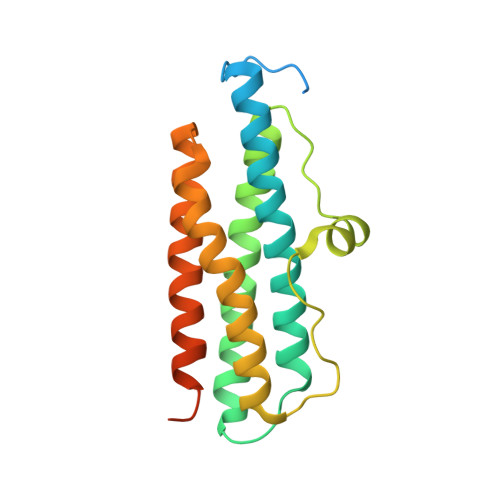
5XGO - PubMed Abstract:
Cage proteins assemble into nanoscale structures with large central cavities. They play roles, including those as virus capsids and chaperones, and have been applied to drug delivery and nanomaterials. Furthermore, protein cages have been used as model systems to understand and design protein quaternary structure. Ferritins are ubiquitous protein cages that manage iron homeostasis and oxidative damage. Two ferritin subfamilies have strongly similar tertiary structure yet distinct quaternary structure: maxi-ferritins normally assemble into 24-meric, octahedral cages with C-terminal E-helices centered around 4-fold symmetry axes, and mini-ferritins are 12-meric, tetrahedral cages with 3-fold axes defined by C-termini lacking E-domains. To understand the role E-domains play in ferritin quaternary structure, we previously designed a chimera of a maxi-ferritin E-domain fused to the C-terminus of a mini-ferritin. The chimera is a 12-mer cage midway in size between those of the maxi- and mini-ferritin. The research described herein sets out to understand (a) whether the increase in size over a typical mini-ferritin is due to a frozen state where the E-domain is flipped out of the cage and (b) whether the symmetrical preference of the E-domain in the maxi-ferritin (4-fold axis) overrules the C-terminal preference in the mini-ferritin (3-fold axis). With a 1.99 Å resolution crystal structure, we determined that the chimera assembles into a tetrahedral cage that can be nearly superimposed with the parent mini-ferritin, and that the E-domains are flipped external to the cage at the 3-fold symmetry axes.
Organizational Affiliation:
Department of Chemistry, King's College London , London, U.K.