5XDO

Crystal structure of human voltage-dependent anion channel 1 (hVDAC1) in C222 space group


Experimental Data Snapshot

  • Method: X-RAY DIFFRACTION
  • Resolution: 3.10 Å
  • R-Value Free: 0.298 
  • R-Value Work: 0.255 
  • R-Value Observed: 0.258 

wwPDB Validation   3D Report Full Report


This is version 1.2 of the entry. See complete history


Literature

Crystal structural characterization reveals novel oligomeric interactions of human voltage-dependent anion channel 1

Hosaka, T.Okazaki, M.Kimura-Someya, T.Ishizuka-Katsura, Y.Ito, K.Yokoyama, S.Dodo, K.Sodeoka, M.Shirouzu, M.

(2017) Protein Sci 26: 1749-1758

  • DOI: https://doi.org/10.1002/pro.3211
  • Primary Citation of Related Structures:  
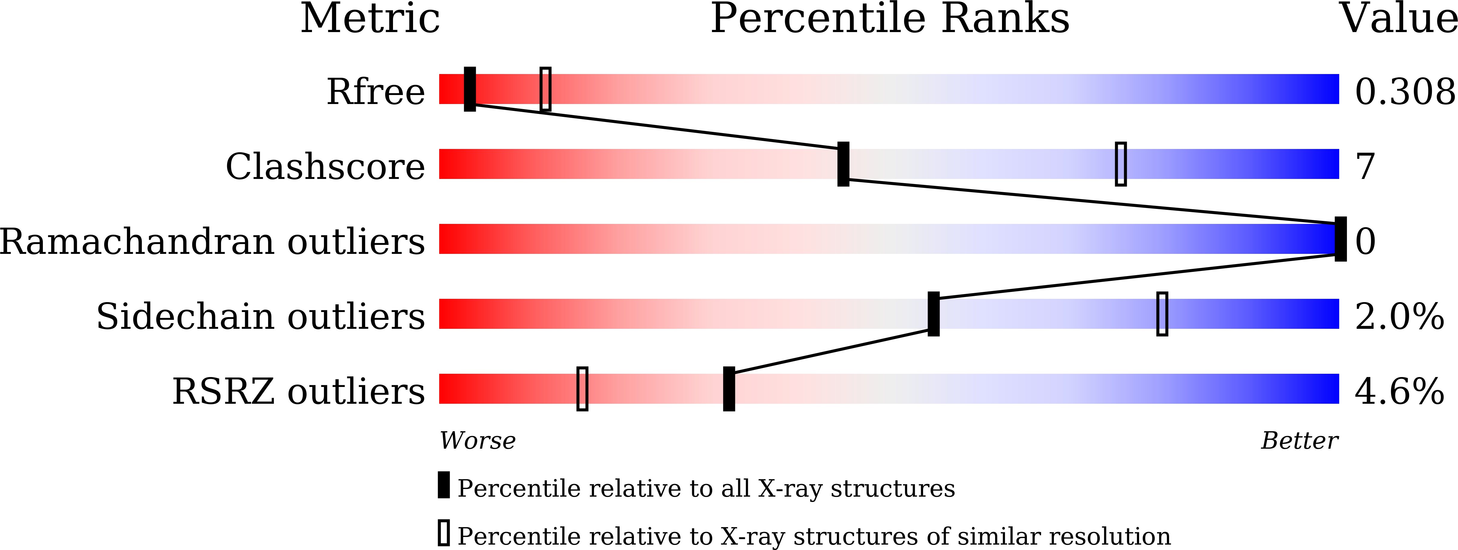
    5XDN, 5XDO

  • PubMed Abstract: 

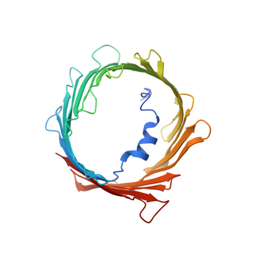
    Voltage-dependent anion channel 1 (VDAC1), which is located in the outer mitochondrial membrane, plays important roles in various cellular processes. For example, oligomerization of VDAC1 is involved in the release of cytochrome c to the cytoplasm, leading to apoptosis. However, it is unknown how VDAC1 oligomerization occurs in the membrane. In the present study, we determined high-resolution crystal structures of oligomeric human VDAC1 (hVDAC1) prepared by using an Escherichia coli cell-free protein synthesis system, which avoided the need for denaturation and refolding of the protein. Broad-range screening using a bicelle crystallization method produced crystals in space groups C222 and P22 1 2 1 , which diffracted to a resolution of 3.10 and 3.15 Å, respectively. Each crystal contained two hVDAC1 protomers in the asymmetric unit. Dimer within the asymmetrical unit of the crystal in space group C222 were oriented parallel, whereas those of the crystal in space group P22 1 2 1 were oriented anti-parallel. From a model of the crystal in space group C222, which we constructed by using crystal symmetry operators, a heptameric structure with eight patterns of interaction between protomers, including hydrophobic interactions with β-strands, hydrophilic interactions with loop regions, and protein-lipid interactions, was observed. It is possible that by having multiple patterns of interaction, VDAC1 can form homo- or hetero-oligomers not only with other VDAC1 protomers but also with other proteins such as VDAC2, VDAC3 and apoptosis-regulating proteins in the Bcl-2 family.


  • Organizational Affiliation

    Division of Structural and Synthetic Biology, RIKEN Center for Life Science Technologies, Yokohama, Kanagawa, 230-0045, Japan.


Macromolecules
Find similar proteins by:  (by identity cutoff)  |  3D Structure
Entity ID: 1
MoleculeChains Sequence LengthOrganismDetailsImage
Voltage-dependent anion-selective channel protein 1
A, B
295Homo sapiensMutation(s): 0 
Gene Names: VDAC1VDAC
Membrane Entity: Yes 
UniProt & NIH Common Fund Data Resources
Find proteins for P21796 (Homo sapiens)
Explore P21796 
Go to UniProtKB:  P21796
PHAROS:  P21796
GTEx:  ENSG00000213585 
Entity Groups  
Sequence Clusters30% Identity50% Identity70% Identity90% Identity95% Identity100% Identity
UniProt GroupP21796
Sequence Annotations
Expand
  • Reference Sequence
Small Molecules
Ligands 4 Unique
IDChains Name / Formula / InChI Key2D Diagram3D Interactions
SPD
Query on SPD

Download Ideal Coordinates CCD File 
C [auth A],
I [auth B]
SPERMIDINE
C7 H19 N3
ATHGHQPFGPMSJY-UHFFFAOYSA-N
OCT
Query on OCT

Download Ideal Coordinates CCD File 
D [auth A],
E [auth A],
J [auth B],
K [auth B]
N-OCTANE
C8 H18
TVMXDCGIABBOFY-UHFFFAOYSA-N
HEX
Query on HEX

Download Ideal Coordinates CCD File 
H [auth A]HEXANE
C6 H14
VLKZOEOYAKHREP-UHFFFAOYSA-N
LNK
Query on LNK

Download Ideal Coordinates CCD File 
F [auth A],
G [auth A]
PENTANE
C5 H12
OFBQJSOFQDEBGM-UHFFFAOYSA-N
Experimental Data & Validation

Experimental Data

  • Method: X-RAY DIFFRACTION
  • Resolution: 3.10 Å
  • R-Value Free: 0.298 
  • R-Value Work: 0.255 
  • R-Value Observed: 0.258 
  • Space Group: C 2 2 2
Unit Cell:
Length ( Å )Angle ( ˚ )
a = 121.62α = 90
b = 146.33β = 90
c = 77.53γ = 90
Software Package:
Software NamePurpose
PHENIXrefinement
XSCALEdata scaling
PDB_EXTRACTdata extraction
XDSdata reduction
PHASERphasing

Structure Validation

View Full Validation Report


Entry History 

Deposition Data

Revision History  (Full details and data files)

  • Version 1.0: 2017-06-28
    Type: Initial release
  • Version 1.1: 2017-09-06
    Changes: Database references
  • Version 1.2: 2023-11-22
    Changes: Data collection, Database references, Refinement description