5XAM

Crystal structure of SecDF in I form at 4 A resolution


Experimental Data Snapshot

  • Method: X-RAY DIFFRACTION
  • Resolution: 4.00 Å
  • R-Value Free: 0.402 
  • R-Value Work: 0.333 
  • R-Value Observed: 0.340 

wwPDB Validation   3D Report Full Report


This is version 1.6 of the entry. See complete history


Literature

Tunnel Formation Inferred from the I-Form Structures of the Proton-Driven Protein Secretion Motor SecDF

Furukawa, A.Yoshikaie, K.Mori, T.Mori, H.Morimoto, Y.V.Sugano, Y.Iwaki, S.Minamino, T.Sugita, Y.Tanaka, Y.Tsukazaki, T.

(2017) Cell Rep 19: 895-901

  • DOI: https://doi.org/10.1016/j.celrep.2017.04.030
  • Primary Citation of Related Structures:  
    5XAM, 5XAN, 5XAP

  • PubMed Abstract: 

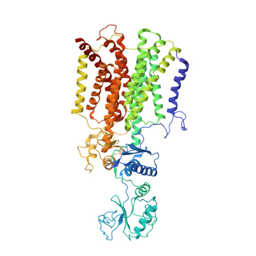
    Protein secretion mediated by SecYEG translocon and SecA ATPase is enhanced by membrane-embedded SecDF by using proton motive force. A previous structural study of SecDF indicated that it comprises 12 transmembrane helices that can conduct protons and three periplasmic domains, which form at least two characterized transition states, termed the F and I forms. We report the structures of full-length SecDF in I form at 2.6- to 2.8-Å resolution. The structures revealed that SecDF in I form can generate a tunnel that penetrates the transmembrane region and functions as a proton pathway regulated by a conserved Asp residue of the transmembrane region. In one crystal structure, periplasmic cavity interacts with a molecule, potentially polyethylene glycol, which may mimic a substrate peptide. This study provides structural insights into the Sec protein translocation that allows future analyses to develop a more detailed working model for SecDF.


  • Organizational Affiliation

    Graduate School of Biological Sciences, Nara Institute of Science and Technology, 8916-5 Takayama-cho, Ikoma, Nara 630-0192, Japan.


Macromolecules
Find similar proteins by:  (by identity cutoff)  |  3D Structure
Entity ID: 1
MoleculeChains Sequence LengthOrganismDetailsImage
Protein translocase subunit SecD
A, B
750Deinococcus radiodurans R1 = ATCC 13939 = DSM 20539Mutation(s): 0 
Gene Names: secDsecFDR_1822
Membrane Entity: Yes 
UniProt
Find proteins for Q9RTE3 (Deinococcus radiodurans (strain ATCC 13939 / DSM 20539 / JCM 16871 / CCUG 27074 / LMG 4051 / NBRC 15346 / NCIMB 9279 / VKM B-1422 / R1))
Explore Q9RTE3 
Go to UniProtKB:  Q9RTE3
Entity Groups  
Sequence Clusters30% Identity50% Identity70% Identity90% Identity95% Identity100% Identity
UniProt GroupQ9RTE3
Sequence Annotations
Expand
  • Reference Sequence
Experimental Data & Validation

Experimental Data

  • Method: X-RAY DIFFRACTION
  • Resolution: 4.00 Å
  • R-Value Free: 0.402 
  • R-Value Work: 0.333 
  • R-Value Observed: 0.340 
  • Space Group: P 1 21 1
Unit Cell:
Length ( Å )Angle ( ˚ )
a = 93.32α = 90
b = 62.26β = 101.71
c = 181.71γ = 90
Software Package:
Software NamePurpose
HKL-2000data collection
SCALEPACKdata scaling
PHASERphasing
PHENIXrefinement
PDB_EXTRACTdata extraction
PHASERphasing
HKLdata reduction

Structure Validation

View Full Validation Report


Entry History & Funding Information

Deposition Data


Funding OrganizationLocationGrant Number
JSPS KAKENHIJapanJP26119006, JP26119007, JP26291023, JP16K14713, JP15H01537, 15H05594, JP15J08235, JP15K06972, JP15K14490

Revision History  (Full details and data files)

  • Version 1.0: 2017-05-17
    Type: Initial release
  • Version 1.1: 2017-10-04
    Changes: Data collection
  • Version 1.2: 2019-05-22
    Changes: Data collection, Structure summary
  • Version 1.3: 2020-10-14
    Changes: Source and taxonomy
  • Version 1.4: 2020-11-11
    Changes: Data collection
  • Version 1.5: 2022-02-09
    Changes: Database references, Structure summary
  • Version 1.6: 2023-11-22
    Changes: Data collection, Refinement description