5VQ4

Nitrogenase Av1 at pH 5


Experimental Data Snapshot

  • Method: X-RAY DIFFRACTION
  • Resolution: 2.30 Å
  • R-Value Free: 0.222 
  • R-Value Work: 0.168 
  • R-Value Observed: 0.170 

wwPDB Validation   3D Report Full Report


Ligand Structure Quality Assessment 


This is version 1.4 of the entry. See complete history


Literature

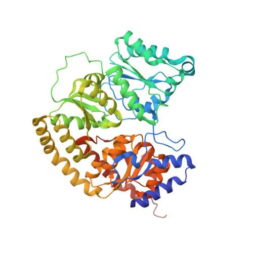
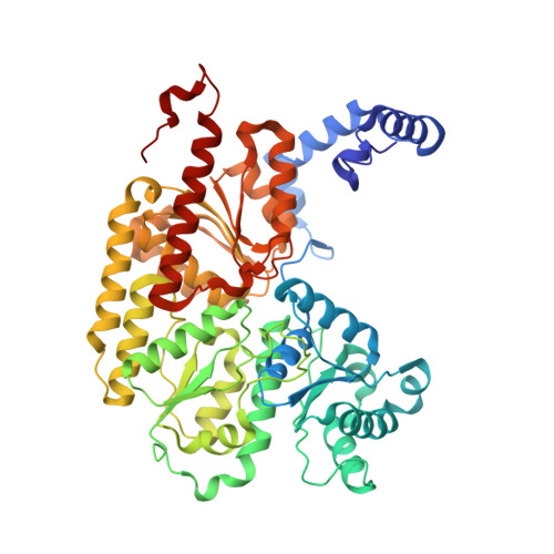
Reversible Protonated Resting State of the Nitrogenase Active Site.

Morrison, C.N.Spatzal, T.Rees, D.C.

(2017) J Am Chem Soc 139: 10856-10862

  • DOI: https://doi.org/10.1021/jacs.7b05695
  • Primary Citation of Related Structures:  
    5VPW, 5VQ3, 5VQ4

  • PubMed Abstract: 

    Protonated states of the nitrogenase active site are mechanistically significant since substrate reduction is invariably accompanied by proton uptake. We report the low pH characterization by X-ray crystallography and EPR spectroscopy of the nitrogenase molybdenum iron (MoFe) proteins from two phylogenetically distinct nitrogenases (Azotobacter vinelandii, Av, and Clostridium pasteurianum, Cp) at pHs between 4.5 and 8. X-ray data at pHs of 4.5-6 reveal the repositioning of side chains along one side of the FeMo-cofactor, and the corresponding EPR data shows a new S = 3/2 spin system with spectral features similar to a state previously observed during catalytic turnover. The structural changes suggest that FeMo-cofactor belt sulfurs S3A or S5A are potential protonation sites. Notably, the observed structural and electronic low pH changes are correlated and reversible. The detailed structural rearrangements differ between the two MoFe proteins, which may reflect differences in potential protonation sites at the active site among nitrogenase species. These observations emphasize the benefits of investigating multiple nitrogenase species. Our experimental data suggest that reversible protonation of the resting state is likely occurring, and we term this state "E 0 H + ", following the Lowe-Thorneley naming scheme.


  • Organizational Affiliation

    Division of Chemistry and Chemical Engineering and ‡Howard Hughes Medical Institute, California Institute of Technology , Pasadena, California 91125, United States.


Macromolecules
Find similar proteins by:  (by identity cutoff)  |  3D Structure
Entity ID: 1
MoleculeChains Sequence LengthOrganismDetailsImage
Nitrogenase molybdenum-iron protein alpha chain
A, C
492Azotobacter vinelandiiMutation(s): 0 
Gene Names: nifD
EC: 1.18.6.1
UniProt
Find proteins for P07328 (Azotobacter vinelandii)
Explore P07328 
Go to UniProtKB:  P07328
Entity Groups  
Sequence Clusters30% Identity50% Identity70% Identity90% Identity95% Identity100% Identity
UniProt GroupP07328
Sequence Annotations
Expand
  • Reference Sequence
Find similar proteins by:  (by identity cutoff)  |  3D Structure
Entity ID: 2
MoleculeChains Sequence LengthOrganismDetailsImage
Nitrogenase molybdenum-iron protein beta chain
B, D
523Azotobacter vinelandiiMutation(s): 0 
Gene Names: nifK
EC: 1.18.6.1
UniProt
Find proteins for P07329 (Azotobacter vinelandii)
Explore P07329 
Go to UniProtKB:  P07329
Entity Groups  
Sequence Clusters30% Identity50% Identity70% Identity90% Identity95% Identity100% Identity
UniProt GroupP07329
Sequence Annotations
Expand
  • Reference Sequence
Small Molecules
Ligands 4 Unique
IDChains Name / Formula / InChI Key2D Diagram3D Interactions
ICS
Query on ICS

Download Ideal Coordinates CCD File 
F [auth A],
K [auth C]
iron-sulfur-molybdenum cluster with interstitial carbon
C Fe7 Mo S9
DDQFAOMIVKLFON-UHFFFAOYSA-N
CLF
Query on CLF

Download Ideal Coordinates CCD File 
G [auth A],
L [auth C]
FE(8)-S(7) CLUSTER
Fe8 S7
JKVMXLBGZBULKV-UHFFFAOYSA-N
HCA
Query on HCA

Download Ideal Coordinates CCD File 
E [auth A],
J [auth C]
3-HYDROXY-3-CARBOXY-ADIPIC ACID
C7 H10 O7
XKJVEVRQMLKSMO-SSDOTTSWSA-N
FE
Query on FE

Download Ideal Coordinates CCD File 
H [auth B],
I [auth B]
FE (III) ION
Fe
VTLYFUHAOXGGBS-UHFFFAOYSA-N
Experimental Data & Validation

Experimental Data

  • Method: X-RAY DIFFRACTION
  • Resolution: 2.30 Å
  • R-Value Free: 0.222 
  • R-Value Work: 0.168 
  • R-Value Observed: 0.170 
  • Space Group: P 1 21 1
Unit Cell:
Length ( Å )Angle ( ˚ )
a = 81.315α = 90
b = 128.859β = 110.89
c = 108.359γ = 90
Software Package:
Software NamePurpose
REFMACrefinement
XDSdata scaling
Aimlessdata scaling
PHASERphasing

Structure Validation

View Full Validation Report


Ligand Structure Quality Assessment 


Entry History & Funding Information

Deposition Data


Funding OrganizationLocationGrant Number
National Institutes of Health/National Institute of General Medical Sciences (NIH/NIGMS)United StatesGM45162

Revision History  (Full details and data files)

  • Version 1.0: 2017-07-26
    Type: Initial release
  • Version 1.1: 2017-08-23
    Changes: Database references
  • Version 1.2: 2017-11-22
    Changes: Refinement description
  • Version 1.3: 2020-01-01
    Changes: Author supporting evidence
  • Version 1.4: 2023-10-04
    Changes: Data collection, Database references, Derived calculations, Refinement description