5VL8

Coordination Chemistry within a Protein Host: Regulation of the Secondary Coordination Sphere


Experimental Data Snapshot

  • Method: X-RAY DIFFRACTION
  • Resolution: 1.70 Å
  • R-Value Free: 0.215 
  • R-Value Work: 0.176 
  • R-Value Observed: 0.178 

wwPDB Validation   3D Report Full Report


Ligand Structure Quality Assessment 


This is version 1.3 of the entry. See complete history


Literature

Coordination chemistry within a protein host: regulation of the secondary coordination sphere.

Mann, S.I.Heinisch, T.Ward, T.R.Borovik, A.S.

(2018) Chem Commun (Camb) 54: 4413-4416

  • DOI: https://doi.org/10.1039/c8cc01931b
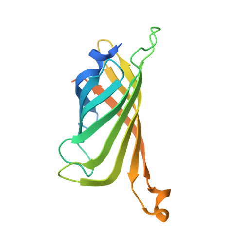
  • Primary Citation of Related Structures:  
    5VKX, 5VL5, 5VL8

  • PubMed Abstract: 

    Secondary coordination spheres of metal complexes are instrumental in controlling properties that are linked to function. To study these effects in aqueous solutions artificial Cu proteins have been developed using biotin-streptavidin (Sav) technology and their binding of external azide ions investigated. Parallel binding studies were done in crystallo on single crystals of the artificial Cu proteins. Spectroscopic changes in solution are consistent with azide binding to the Cu centers. Structural studies corroborate that a Cu-N3 unit is present in each Sav subunit and reveal the composition of hydrogen bonding (H-bonding) networks that include the coordinated azido ligand. The networks involve amino acid residues and water molecules within the secondary coordination sphere. Mutation of these residues to ones that cannot form H-bonds caused a measurble change in the equilibrium binding constants that were measured in solution. These findings further demonstrate the utility of biotin-Sav technology to prepare water-stable inorganic complexes whose structures can be controlled within both primary and secondary coordination spheres.


  • Organizational Affiliation

    Department of Chemistry, 1102 Natural Science II, University of California, Irvine, CA 92697, USA. aborovik@uci.edu.


Macromolecules
Find similar proteins by:  (by identity cutoff)  |  3D Structure
Entity ID: 1
MoleculeChains Sequence LengthOrganismDetailsImage
Streptavidin159Streptomyces avidiniiMutation(s): 0 
UniProt
Find proteins for P22629 (Streptomyces avidinii)
Explore P22629 
Go to UniProtKB:  P22629
Entity Groups  
Sequence Clusters30% Identity50% Identity70% Identity90% Identity95% Identity100% Identity
UniProt GroupP22629
Sequence Annotations
Expand
  • Reference Sequence
Small Molecules
Ligands 3 Unique
IDChains Name / Formula / InChI Key2D Diagram3D Interactions
S32
Query on S32

Download Ideal Coordinates CCD File 
B [auth A][N-(3-{bis[2-(pyridin-2-yl-kappaN)ethyl]amino-kappaN}propyl)-5-(2-oxohexahydro-1H-thieno[3,4-d]imidazol-4-yl)pentanamide](hydroxy)copper
C27 H38 Cu N6 O3 S
PNZAMFOPABFOOE-KNJZPGARSA-M
GOL
Query on GOL

Download Ideal Coordinates CCD File 
D [auth A]GLYCEROL
C3 H8 O3
PEDCQBHIVMGVHV-UHFFFAOYSA-N
CU
Query on CU

Download Ideal Coordinates CCD File 
C [auth A]COPPER (II) ION
Cu
JPVYNHNXODAKFH-UHFFFAOYSA-N
Experimental Data & Validation

Experimental Data

  • Method: X-RAY DIFFRACTION
  • Resolution: 1.70 Å
  • R-Value Free: 0.215 
  • R-Value Work: 0.176 
  • R-Value Observed: 0.178 
  • Space Group: I 41 2 2
Unit Cell:
Length ( Å )Angle ( ˚ )
a = 57.58α = 90
b = 57.58β = 90
c = 183.57γ = 90
Software Package:
Software NamePurpose
REFMACrefinement
iMOSFLMdata reduction
Aimlessdata scaling
PHASERphasing

Structure Validation

View Full Validation Report


Ligand Structure Quality Assessment 


Entry History 

Deposition Data

Revision History  (Full details and data files)

  • Version 1.0: 2018-04-18
    Type: Initial release
  • Version 1.1: 2018-04-25
    Changes: Data collection, Database references
  • Version 1.2: 2018-05-09
    Changes: Data collection, Database references
  • Version 1.3: 2023-10-04
    Changes: Data collection, Database references, Refinement description