5TYH

PglD from Campylobacter jejuni NCTC 11168 in complex with 5-(2-furanyl)-1H-pyrazole-3-carboxylic acid


Experimental Data Snapshot

  • Method: X-RAY DIFFRACTION
  • Resolution: 2.10 Å
  • R-Value Free: 0.241 
  • R-Value Work: 0.196 
  • R-Value Observed: 0.198 

wwPDB Validation   3D Report Full Report


Ligand Structure Quality Assessment 


This is version 1.1 of the entry. See complete history


Literature

Targeting Bacillosamine Biosynthesis in Bacterial Pathogens: Development of Inhibitors to a Bacterial Amino-Sugar Acetyltransferase from Campylobacter jejuni.

De Schutter, J.W.Morrison, J.P.Morrison, M.J.Ciulli, A.Imperiali, B.

(2017) J Med Chem 60: 2099-2118

  • DOI: https://doi.org/10.1021/acs.jmedchem.6b01869
  • Primary Citation of Related Structures:  
    5T2Y, 5TYH

  • PubMed Abstract: 

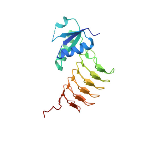
    The glycoproteins of selected microbial pathogens often include highly modified carbohydrates such as 2,4-diacetamidobacillosamine (diNAcBac). These glycoconjugates are involved in host-cell interactions and may be associated with the virulence of medically significant Gram-negative bacteria. In light of genetic studies demonstrating the attenuated virulence of bacterial strains in which modified carbohydrate biosynthesis enzymes have been knocked out, we are developing small molecule inhibitors of selected enzymes as tools to evaluate whether such compounds modulate virulence. We performed fragment-based and high-throughput screens against an amino-sugar acetyltransferase enzyme, PglD, involved in biosynthesis of UDP-diNAcBac in Campylobacter jejuni. Herein we report optimization of the hits into potent small molecule inhibitors (IC 50 < 300 nM). Biophysical characterization shows that the best inhibitors are competitive with acetyl coenzyme A and an X-ray cocrystal structure reveals that binding is biased toward occupation of the adenine subpocket of the AcCoA binding site by an aromatic ring.


  • Organizational Affiliation

    Department of Chemistry, Massachusetts Institute of Technology , 77 Massachusetts Avenue, Cambridge, Massachusetts 02139, United States.


Macromolecules
Find similar proteins by:  (by identity cutoff)  |  3D Structure
Entity ID: 1
MoleculeChains Sequence LengthOrganismDetailsImage
UDP-N-acetylbacillosamine N-acetyltransferase198Campylobacter jejuni subsp. jejuni NCTC 11168 = ATCC 700819Mutation(s): 0 
Gene Names: pglDCj1123c
EC: 2.3.1.203
UniProt
Find proteins for Q0P9D1 (Campylobacter jejuni subsp. jejuni serotype O:2 (strain ATCC 700819 / NCTC 11168))
Explore Q0P9D1 
Go to UniProtKB:  Q0P9D1
Entity Groups  
Sequence Clusters30% Identity50% Identity70% Identity90% Identity95% Identity100% Identity
UniProt GroupQ0P9D1
Sequence Annotations
Expand
  • Reference Sequence
Small Molecules
Ligands 1 Unique
IDChains Name / Formula / InChI Key2D Diagram3D Interactions
7O4
Query on 7O4

Download Ideal Coordinates CCD File 
B [auth A],
C [auth A]
3-(furan-2-yl)-1H-pyrazole-5-carboxylic acid
C8 H6 N2 O3
GKPSFQIKCROJOB-UHFFFAOYSA-N
Binding Affinity Annotations 
IDSourceBinding Affinity
7O4 Binding MOAD:  5TYH IC50: 8.40e+6 (nM) from 1 assay(s)
Experimental Data & Validation

Experimental Data

  • Method: X-RAY DIFFRACTION
  • Resolution: 2.10 Å
  • R-Value Free: 0.241 
  • R-Value Work: 0.196 
  • R-Value Observed: 0.198 
  • Space Group: P 63
Unit Cell:
Length ( Å )Angle ( ˚ )
a = 86.726α = 90
b = 86.726β = 90
c = 65.53γ = 120
Software Package:
Software NamePurpose
REFMACrefinement
MOLREPphasing
SCALEPACKdata scaling
PHASERphasing
HKL-2000data reduction

Structure Validation

View Full Validation Report


Ligand Structure Quality Assessment 


Entry History 

Deposition Data

Revision History  (Full details and data files)

  • Version 1.0: 2017-04-12
    Type: Initial release
  • Version 1.1: 2023-10-04
    Changes: Data collection, Database references, Refinement description