5TV1

active arrestin-3 with inositol hexakisphosphate


Experimental Data Snapshot

  • Method: X-RAY DIFFRACTION
  • Resolution: 2.40 Å
  • R-Value Free: 0.245 
  • R-Value Work: 0.197 
  • R-Value Observed: 0.199 

wwPDB Validation   3D Report Full Report


Ligand Structure Quality Assessment 


This is version 1.3 of the entry. See complete history


Literature

Structural basis of arrestin-3 activation and signaling.

Chen, Q.Perry, N.A.Vishnivetskiy, S.A.Berndt, S.Gilbert, N.C.Zhuo, Y.Singh, P.K.Tholen, J.Ohi, M.D.Gurevich, E.V.Brautigam, C.A.Klug, C.S.Gurevich, V.V.Iverson, T.M.

(2017) Nat Commun 8: 1427-1427

  • DOI: https://doi.org/10.1038/s41467-017-01218-8
  • Primary Citation of Related Structures:  
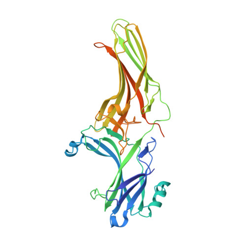
    5TV1

  • PubMed Abstract: 

    A unique aspect of arrestin-3 is its ability to support both receptor-dependent and receptor-independent signaling. Here, we show that inositol hexakisphosphate (IP 6 ) is a non-receptor activator of arrestin-3 and report the structure of IP 6 -activated arrestin-3 at 2.4-Å resolution. IP 6 -activated arrestin-3 exhibits an inter-domain twist and a displaced C-tail, hallmarks of active arrestin. IP 6 binds to the arrestin phosphate sensor, and is stabilized by trimerization. Analysis of the trimerization surface, which is also the receptor-binding surface, suggests a feature called the finger loop as a key region of the activation sensor. We show that finger loop helicity and flexibility may underlie coupling to hundreds of diverse receptors and also promote arrestin-3 activation by IP 6 . Importantly, we show that effector-binding sites on arrestins have distinct conformations in the basal and activated states, acting as switch regions. These switch regions may work with the inter-domain twist to initiate and direct arrestin-mediated signaling.


  • Organizational Affiliation

    Department of Pharmacology, Vanderbilt University, Nashville, TN, 37232, USA.


Macromolecules
Find similar proteins by:  (by identity cutoff)  |  3D Structure
Entity ID: 1
MoleculeChains Sequence LengthOrganismDetailsImage
Beta-arrestin-2393Bos taurusMutation(s): 0 
Gene Names: ARRB2
UniProt
Find proteins for P32120 (Bos taurus)
Explore P32120 
Go to UniProtKB:  P32120
Entity Groups  
Sequence Clusters30% Identity50% Identity70% Identity90% Identity95% Identity100% Identity
UniProt GroupP32120
Sequence Annotations
Expand
  • Reference Sequence
Experimental Data & Validation

Experimental Data

  • Method: X-RAY DIFFRACTION
  • Resolution: 2.40 Å
  • R-Value Free: 0.245 
  • R-Value Work: 0.197 
  • R-Value Observed: 0.199 
  • Space Group: P 63
Unit Cell:
Length ( Å )Angle ( ˚ )
a = 97.575α = 90
b = 97.575β = 90
c = 76.938γ = 120
Software Package:
Software NamePurpose
PHENIXrefinement
HKL-2000data scaling
HKL-2000data reduction
PDB_EXTRACTdata extraction
PHASERphasing

Structure Validation

View Full Validation Report


Ligand Structure Quality Assessment 


Entry History & Funding Information

Deposition Data


Funding OrganizationLocationGrant Number
National Institutes of Health/National Institute of General Medical Sciences (NIH/NIGMS)United StatesGM095633
National Institutes of Health/National Institute of General Medical Sciences (NIH/NIGMS)United StatesGM077561
National Institutes of Health/National Institute of General Medical Sciences (NIH/NIGMS)United StatesGM109955
National Institutes of Health/National Eye Institute (NIH/NEI)United StatesEY011500

Revision History  (Full details and data files)

  • Version 1.0: 2017-11-22
    Type: Initial release
  • Version 1.1: 2019-12-11
    Changes: Author supporting evidence
  • Version 1.2: 2020-10-14
    Changes: Structure summary
  • Version 1.3: 2023-10-04
    Changes: Data collection, Database references, Refinement description