5THP

Rhodocetin in complex with the integrin alpha2-A domain


Experimental Data Snapshot

  • Method: X-RAY DIFFRACTION
  • Resolution: 3.01 Å
  • R-Value Free: 0.270 
  • R-Value Work: 0.221 
  • R-Value Observed: 0.224 

wwPDB Validation   3D Report Full Report


This is version 1.1 of the entry. See complete history


Literature

Dramatic and concerted conformational changes enable rhodocetin to block alpha 2 beta 1 integrin selectively.

Eble, J.A.McDougall, M.Orriss, G.L.Niland, S.Johanningmeier, B.Pohlentz, G.Meier, M.Karrasch, S.Estevao-Costa, M.I.Martins Lima, A.Stetefeld, J.

(2017) PLoS Biol 15: e2001492-e2001492

  • DOI: https://doi.org/10.1371/journal.pbio.2001492
  • Primary Citation of Related Structures:  
    5THP

  • PubMed Abstract: 

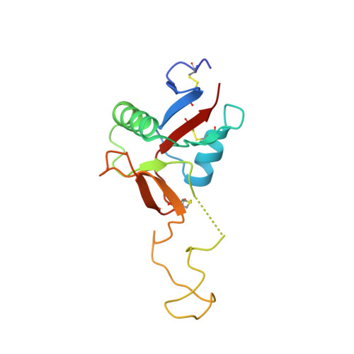
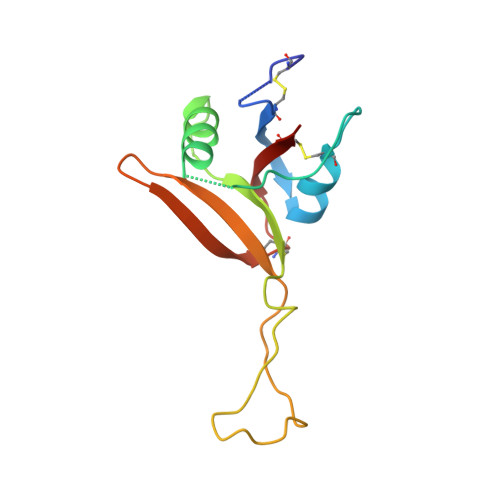
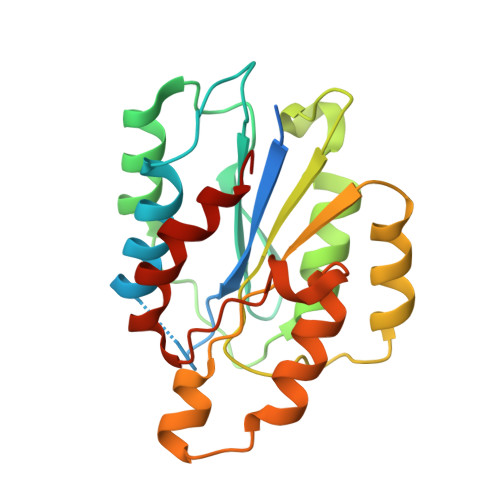
    The collagen binding integrin α2β1 plays a crucial role in hemostasis, fibrosis, and cancer progression amongst others. It is specifically inhibited by rhodocetin (RC), a C-type lectin-related protein (CLRP) found in Malayan pit viper (Calloselasma rhodostoma) venom. The structure of RC alone reveals a heterotetramer arranged as an αβ and γδ subunit in a cruciform shape. RC specifically binds to the collagen binding A-domain of the integrin α2 subunit, thereby blocking collagen-induced platelet aggregation. However, until now, the molecular basis for this interaction has remained unclear. Here, we present the molecular structure of the RCγδ-α2A complex solved to 3.0 Å resolution. Our findings show that RC undergoes a dramatic structural reorganization upon binding to α2β1 integrin. Besides the release of the nonbinding RCαβ tandem, the RCγ subunit interacts with loop 2 of the α2A domain as result of a dramatic conformational change. The RCδ subunit contacts the integrin α2A domain in the "closed" conformation through its helix C. Combined with epitope-mapped antibodies, conformationally locked α2A domain mutants, point mutations within the α2A loop 2, and chemical modifications of the purified toxin protein, this molecular structure of RCγδ-α2A complex explains the inhibitory mechanism and specificity of RC for α2β1 integrin.


  • Organizational Affiliation

    Institute of Physiological Chemistry and Pathobiochemistry, University of Münster, Münster, Germany.


Macromolecules
Find similar proteins by:  (by identity cutoff)  |  3D Structure
Entity ID: 1
MoleculeChains Sequence LengthOrganismDetailsImage
Snaclec rhodocetin subunit gamma
A, D, G, J, M
A, D, G, J, M, P
135Calloselasma rhodostomaMutation(s): 0 
UniProt
Find proteins for D2YW39 (Calloselasma rhodostoma)
Explore D2YW39 
Go to UniProtKB:  D2YW39
Entity Groups  
Sequence Clusters30% Identity50% Identity70% Identity90% Identity95% Identity100% Identity
UniProt GroupD2YW39
Sequence Annotations
Expand
  • Reference Sequence
Find similar proteins by:  (by identity cutoff)  |  3D Structure
Entity ID: 2
MoleculeChains Sequence LengthOrganismDetailsImage
Snaclec rhodocetin subunit delta
B, E, H, K, N
B, E, H, K, N, Q
124Calloselasma rhodostomaMutation(s): 0 
UniProt
Find proteins for D2YW40 (Calloselasma rhodostoma)
Explore D2YW40 
Go to UniProtKB:  D2YW40
Entity Groups  
Sequence Clusters30% Identity50% Identity70% Identity90% Identity95% Identity100% Identity
UniProt GroupD2YW40
Sequence Annotations
Expand
  • Reference Sequence
Find similar proteins by:  (by identity cutoff)  |  3D Structure
Entity ID: 3
MoleculeChains Sequence LengthOrganismDetailsImage
Integrin alpha-2
C, F, I, L, O
C, F, I, L, O, R
217Homo sapiensMutation(s): 0 
Gene Names: ITGA2CD49B
UniProt & NIH Common Fund Data Resources
Find proteins for P17301 (Homo sapiens)
Explore P17301 
Go to UniProtKB:  P17301
PHAROS:  P17301
GTEx:  ENSG00000164171 
Entity Groups  
Sequence Clusters30% Identity50% Identity70% Identity90% Identity95% Identity100% Identity
UniProt GroupP17301
Sequence Annotations
Expand
  • Reference Sequence
Small Molecules
Ligands 5 Unique
IDChains Name / Formula / InChI Key2D Diagram3D Interactions
SO4
Query on SO4

Download Ideal Coordinates CCD File 
CB [auth F]
DC [auth L]
DD [auth R]
EA [auth C]
MC [auth O]
CB [auth F],
DC [auth L],
DD [auth R],
EA [auth C],
MC [auth O],
OA [auth D],
RC [auth O],
SB [auth I],
WA [auth E],
XA [auth F]
SULFATE ION
O4 S
QAOWNCQODCNURD-UHFFFAOYSA-L
GOL
Query on GOL

Download Ideal Coordinates CCD File 
FA [auth C]
GB [auth G]
HB [auth G]
LB [auth H]
U [auth A]
FA [auth C],
GB [auth G],
HB [auth G],
LB [auth H],
U [auth A],
VC [auth P],
WB [auth J],
XB [auth J]
GLYCEROL
C3 H8 O3
PEDCQBHIVMGVHV-UHFFFAOYSA-N
CL
Query on CL

Download Ideal Coordinates CCD File 
AA [auth C]
BA [auth C]
BB [auth F]
BC [auth L]
CA [auth C]
AA [auth C],
BA [auth C],
BB [auth F],
BC [auth L],
CA [auth C],
CC [auth L],
CD [auth R],
DA [auth C],
EB [auth G],
FB [auth G],
FC [auth M],
GC [auth M],
IC [auth N],
JA [auth D],
JB [auth H],
JC [auth N],
KA [auth D],
KB [auth H],
KC [auth N],
LA [auth D],
MA [auth D],
NA [auth D],
PB [auth I],
QB [auth I],
QC [auth O],
RB [auth I],
T [auth A],
TA [auth E],
TC [auth P],
UA [auth E],
UC [auth P],
VA [auth E],
VB [auth J],
YC [auth Q],
ZC [auth Q]
CHLORIDE ION
Cl
VEXZGXHMUGYJMC-UHFFFAOYSA-M
MG
Query on MG

Download Ideal Coordinates CCD File 
AB [auth F]
AC [auth L]
BD [auth R]
OB [auth I]
PC [auth O]
AB [auth F],
AC [auth L],
BD [auth R],
OB [auth I],
PC [auth O],
Z [auth C]
MAGNESIUM ION
Mg
JLVVSXFLKOJNIY-UHFFFAOYSA-N
NA
Query on NA

Download Ideal Coordinates CCD File 
AD [auth R]
DB [auth G]
EC [auth M]
GA [auth C]
HA [auth D]
AD [auth R],
DB [auth G],
EC [auth M],
GA [auth C],
HA [auth D],
HC [auth N],
IA [auth D],
IB [auth H],
LC [auth O],
MB [auth I],
NB [auth I],
NC [auth O],
OC [auth O],
PA [auth D],
QA [auth E],
RA [auth E],
S [auth A],
SA [auth E],
SC [auth P],
TB [auth J],
UB [auth J],
V [auth B],
W [auth C],
WC [auth Q],
X [auth C],
XC [auth Q],
Y [auth C],
YA [auth F],
YB [auth L],
ZA [auth F],
ZB [auth L]
SODIUM ION
Na
FKNQFGJONOIPTF-UHFFFAOYSA-N
Experimental Data & Validation

Experimental Data

  • Method: X-RAY DIFFRACTION
  • Resolution: 3.01 Å
  • R-Value Free: 0.270 
  • R-Value Work: 0.221 
  • R-Value Observed: 0.224 
  • Space Group: P 41
Unit Cell:
Length ( Å )Angle ( ˚ )
a = 130.763α = 90
b = 130.763β = 90
c = 251.351γ = 90
Software Package:
Software NamePurpose
PHENIXrefinement
XDSdata reduction
Aimlessdata scaling
PHASERphasing

Structure Validation

View Full Validation Report


Entry History & Funding Information

Deposition Data


Funding OrganizationLocationGrant Number
Heart and Stroke Foundation Canada, Grant-in-Aid programCanadaG-14-0006025

Revision History  (Full details and data files)

  • Version 1.0: 2017-08-02
    Type: Initial release
  • Version 1.1: 2023-10-04
    Changes: Data collection, Database references, Derived calculations, Refinement description