Photorhabdus luminescens lectin A (PllA): A new probe for detecting alpha-galactoside-terminating glycoconjugates.
Beshr, G., Sikandar, A., Jemiller, E.M., Klymiuk, N., Hauck, D., Wagner, S., Wolf, E., Koehnke, J., Titz, A.(2017) J Biol Chem 292: 19935-19951
- PubMed: 28972138
- DOI: https://doi.org/10.1074/jbc.M117.812792
- Primary Citation of Related Structures:
5ODU, 5OFI, 5OFX, 5OFZ - PubMed Abstract:
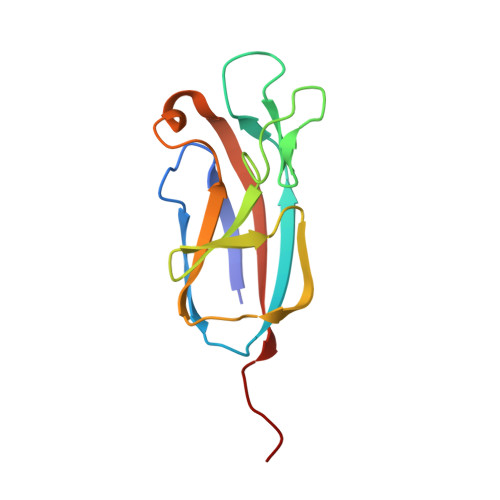
Lectins play important roles in infections by pathogenic bacteria, for example, in host colonization, persistence, and biofilm formation. The Gram-negative entomopathogenic bacterium Photorhabdus luminescens symbiotically lives in insect-infecting Heterorhabditis nematodes and kills the insect host upon invasion by the nematode. The P. luminescens genome harbors the gene plu2096 , coding for a novel lectin that we named PllA. We analyzed the binding properties of purified PllA with a glycan array and a binding assay in solution. Both assays revealed a strict specificity of PllA for α-galactoside-terminating glycoconjugates. The crystal structures of apo PllA and complexes with three different ligands revealed the molecular basis for the strict specificity of this lectin. Furthermore, we found that a 90° twist in subunit orientation leads to a peculiar quaternary structure compared with that of its ortholog LecA from Pseudomonas aeruginosa We also investigated the utility of PllA as a probe for detecting α-galactosides. The α-Gal epitope is present on wild-type pig cells and is the main reason for hyperacute organ rejection in pig to primate xenotransplantation. We noted that PllA specifically recognizes this epitope on the glycan array and demonstrated that PllA can be used as a fluorescent probe to detect this epitope on primary porcine cells in vitro In summary, our biochemical and structural analyses of the P. luminescens lectin PllA have disclosed the structural basis for PllA's high specificity for α-galactoside-containing ligands, and we show that PllA can be used to visualize the α-Gal epitope on porcine tissues.
Organizational Affiliation:
From the Divisions of Chemical Biology of Carbohydrates and.