5OBA

Structure of a modified mouse H-chain ferritin with a lanthanide binding motif


Experimental Data Snapshot

  • Method: X-RAY DIFFRACTION
  • Resolution: 2.85 Å
  • R-Value Free: 0.170 
  • R-Value Work: 0.137 
  • R-Value Observed: 0.138 

wwPDB Validation   3D Report Full Report


This is version 1.2 of the entry. See complete history


Literature

Engineered ferritin for lanthanide binding.

Calisti, L.Trabuco, M.C.Boffi, A.Testi, C.Montemiglio, L.C.des Georges, A.Benni, I.Ilari, A.Taciak, B.Bialasek, M.Rygiel, T.Krol, M.Baiocco, P.Bonamore, A.

(2018) PLoS One 13: e0201859-e0201859

  • DOI: https://doi.org/10.1371/journal.pone.0201859
  • Primary Citation of Related Structures:  
    5OBA, 5OBB

  • PubMed Abstract: 

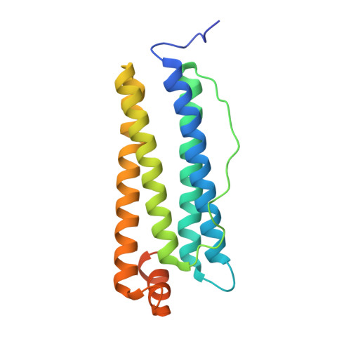
    Ferritin H-homopolymers have been extensively used as nanocarriers for diverse applications in the targeted delivery of drugs and imaging agents, due to their unique ability to bind the transferrin receptor (CD71), highly overexpressed in most tumor cells. In order to incorporate novel fluorescence imaging properties, we have fused a lanthanide binding tag (LBT) to the C-terminal end of mouse H-chain ferritin, HFt. The HFt-LBT possesses one high affinity Terbium binding site per each of the 24 subunits provided by six coordinating aminoacid side chains and a tryptophan residue in its close proximity and is thus endowed with strong FRET sensitization properties. Accordingly, the characteristic Terbium emission band at 544 nm for the HFt-LBT Tb(III) complex was detectable upon excitation of the tag enclosed at two order of magnitude higher intensity with respect to the wtHFt protein. X-ray data at 2.9 Å and cryo-EM at 7 Å resolution demonstrated that HFt-LBT is correctly assembled as a 24-mer both in crystal and in solution. On the basis of the intrinsic Tb(III) binding properties of the wt protein, 32 additional Tb(III) binding sites, located within the natural iron binding sites of the protein, were identified besides the 24 Tb(III) ions coordinated to the LBTs. HFt-LBT Tb(III) was demonstrated to be actively uptaken by selected tumor cell lines by confocal microscopy and FACS analysis of their FITC derivatives, although direct fluorescence from Terbium emission could not be singled out with conventional, 295-375 nm, fluorescence excitation.


  • Organizational Affiliation

    Department of Biochemical Sciences "Alessandro Rossi Fanelli", Sapienza University of Rome, Rome Italy.


Macromolecules
Find similar proteins by:  (by identity cutoff)  |  3D Structure
Entity ID: 1
MoleculeChains Sequence LengthOrganismDetailsImage
Ferritin heavy chain
A, B, C, D, E
A, B, C, D, E, F, G, H, I, J, K, L, M, N, O, P, Q, R, S, T, U, V, W, X
197Mus musculusMutation(s): 0 
Gene Names: Fth1Fth
EC: 1.16.3.1
UniProt & NIH Common Fund Data Resources
Find proteins for P09528 (Mus musculus)
Explore P09528 
Go to UniProtKB:  P09528
IMPC:  MGI:95588
Entity Groups  
Sequence Clusters30% Identity50% Identity70% Identity90% Identity95% Identity100% Identity
UniProt GroupP09528
Sequence Annotations
Expand
  • Reference Sequence
Small Molecules
Ligands 1 Unique
IDChains Name / Formula / InChI Key2D Diagram3D Interactions
FE
Query on FE

Download Ideal Coordinates CCD File 
AA [auth B]
AB [auth V]
BA [auth C]
BB [auth V]
CA [auth D]
AA [auth B],
AB [auth V],
BA [auth C],
BB [auth V],
CA [auth D],
CB [auth W],
DA [auth E],
DB [auth X],
EA [auth E],
FA [auth F],
GA [auth G],
HA [auth G],
IA [auth H],
JA [auth I],
KA [auth J],
LA [auth K],
MA [auth K],
NA [auth L],
OA [auth M],
PA [auth M],
QA [auth N],
RA [auth O],
SA [auth P],
TA [auth P],
UA [auth Q],
VA [auth R],
WA [auth S],
XA [auth T],
Y [auth A],
YA [auth T],
Z [auth A],
ZA [auth U]
FE (III) ION
Fe
VTLYFUHAOXGGBS-UHFFFAOYSA-N
Experimental Data & Validation

Experimental Data

  • Method: X-RAY DIFFRACTION
  • Resolution: 2.85 Å
  • R-Value Free: 0.170 
  • R-Value Work: 0.137 
  • R-Value Observed: 0.138 
  • Space Group: I 2 2 2
Unit Cell:
Length ( Å )Angle ( ˚ )
a = 237.306α = 90
b = 237.468β = 90
c = 237.527γ = 90
Software Package:
Software NamePurpose
REFMACrefinement
Aimlessdata scaling
PDB_EXTRACTdata extraction
XDSdata reduction
PHASERphasing

Structure Validation

View Full Validation Report


Entry History & Funding Information

Deposition Data


Funding OrganizationLocationGrant Number
European UnionItaly637295

Revision History  (Full details and data files)

  • Version 1.0: 2018-07-25
    Type: Initial release
  • Version 1.1: 2018-08-22
    Changes: Data collection, Database references
  • Version 1.2: 2024-01-17
    Changes: Data collection, Database references, Derived calculations, Refinement description