Heightened Dynamics of the Oxidized Y48H Variant of Human Cytochrome c Increases Its Peroxidatic Activity.
Deacon, O.M., Karsisiotis, A.I., Moreno-Chicano, T., Hough, M.A., Macdonald, C., Blumenschein, T.M.A., Wilson, M.T., Moore, G.R., Worrall, J.A.R.(2017) Biochemistry 56: 6111-6124
- PubMed: 29083920
- DOI: https://doi.org/10.1021/acs.biochem.7b00890
- Primary Citation of Related Structures:
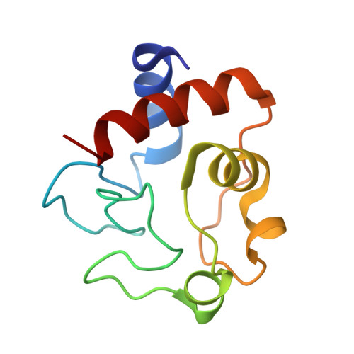
5O10 - PubMed Abstract:
Proteins performing multiple biochemical functions are called "moonlighting proteins" or extreme multifunctional (EMF) proteins. Mitochondrial cytochrome c is an EMF protein that binds multiple partner proteins to act as a signaling molecule, transfers electrons in the respiratory chain, and acts as a peroxidase in apoptosis. Mutations in the cytochrome c gene lead to the disease thrombocytopenia, which is accompanied by enhanced apoptotic activity. The Y48H variant arises from one such mutation and is found in the 40-57 Ω-loop, the lowest-unfolding free energy substructure of the cytochrome c fold. A 1.36 Å resolution X-ray structure of the Y48H variant reveals minimal structural changes compared to the wild-type structure, with the axial Met80 ligand coordinated to the heme iron. Despite this, the intrinsic peroxidase activity is enhanced, implying that a pentacoordinate heme state is more prevalent in the Y48H variant, corroborated through determination of a Met80 "off rate" of >125 s -1 compared to a rate of ∼6 s -1 for the wild-type protein. Heteronuclear nuclear magnetic resonance measurements with the oxidized Y48H variant reveal heightened dynamics in the 40-57 Ω-loop and the Met80-containing 71-85 Ω-loop relative to the wild-type protein, illustrating communication between these substructures. Placed into context with the G41S cytochrome c variant, also implicated in thrombocytopenia, a dynamic picture associated with this disease relative to cytochrome c is emerging whereby increasing dynamics in substructures of the cytochrome c fold serve to facilitate an increased population of the peroxidatic pentacoordinate heme state in the following order: wild type < G41S < Y48H.
Organizational Affiliation:
School of Biological Sciences, University of Essex , Wivenhoe Park, Colchester CO4 3SQ, U.K.