5MN9

Crystal structure of MINDY-1 tMIU in complex with K48-diUb


Experimental Data Snapshot

  • Method: X-RAY DIFFRACTION
  • Resolution: 2.05 Å
  • R-Value Free: 0.223 
  • R-Value Work: 0.183 
  • R-Value Observed: 0.185 

wwPDB Validation   3D Report Full Report


This is version 1.4 of the entry. See complete history


Literature

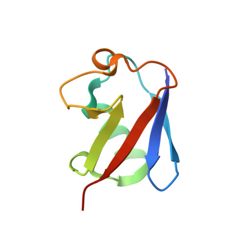
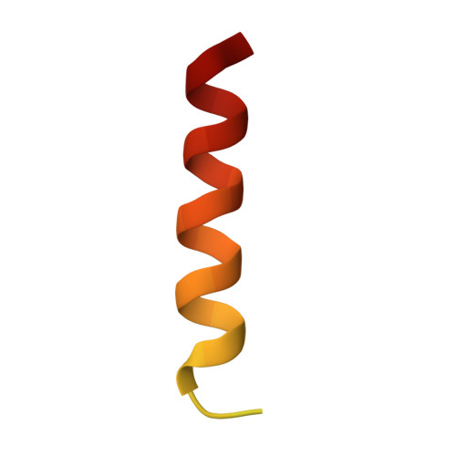
A single MIU motif of MINDY-1 recognizes K48-linked polyubiquitin chains.

Kristariyanto, Y.A.Abdul Rehman, S.A.Weidlich, S.Knebel, A.Kulathu, Y.

(2017) EMBO Rep 18: 392-402

  • DOI: https://doi.org/10.15252/embr.201643205
  • Primary Citation of Related Structures:  
    5MN9

  • PubMed Abstract: 

    The eight different types of ubiquitin (Ub) chains that can be formed play important roles in diverse cellular processes. Linkage-selective recognition of Ub chains by Ub-binding domain (UBD)-containing proteins is central to coupling different Ub signals to specific cellular responses. The motif interacting with ubiquitin (MIU) is a small UBD that has been characterized for its binding to monoUb. The recently discovered deubiquitinase MINDY-1/FAM63A contains a tandem MIU repeat (tMIU) that is highly selective at binding to K48-linked polyUb. We here identify that this linkage-selective binding is mediated by a single MIU motif (MIU2) in MINDY-1. The crystal structure of MIU2 in complex with K48-linked polyubiquitin chains reveals that MIU2 on its own binds to all three Ub moieties in an open conformation that can only be accommodated by K48-linked triUb. The weak Ub binder MIU1 increases overall affinity of the tMIU for polyUb chains without affecting its linkage selectivity. Our analyses reveal new concepts for linkage selectivity and polyUb recognition by UBDs.


  • Organizational Affiliation

    MRC Protein Phosphorylation and Ubiquitylation Unit, School of Life Sciences, University of Dundee, Dundee, UK.


Macromolecules
Find similar proteins by:  (by identity cutoff)  |  3D Structure
Entity ID: 1
MoleculeChains Sequence LengthOrganismDetailsImage
Ubiquitin-40S ribosomal protein S27a
A, B
76Bos taurusMutation(s): 0 
UniProt
Find proteins for P62992 (Bos taurus)
Explore P62992 
Go to UniProtKB:  P62992
Entity Groups  
Sequence Clusters30% Identity50% Identity70% Identity90% Identity95% Identity100% Identity
UniProt GroupP62992
Sequence Annotations
Expand
  • Reference Sequence
Find similar proteins by:  (by identity cutoff)  |  3D Structure
Entity ID: 2
MoleculeChains Sequence LengthOrganismDetailsImage
Ubiquitin carboxyl-terminal hydrolase MINDY-144Homo sapiensMutation(s): 0 
Gene Names: FAM63AKIAA1390
EC: 3.4.19.12
UniProt & NIH Common Fund Data Resources
Find proteins for Q8N5J2 (Homo sapiens)
Explore Q8N5J2 
Go to UniProtKB:  Q8N5J2
PHAROS:  Q8N5J2
GTEx:  ENSG00000143409 
Entity Groups  
Sequence Clusters30% Identity50% Identity70% Identity90% Identity95% Identity100% Identity
UniProt GroupQ8N5J2
Sequence Annotations
Expand
  • Reference Sequence
Experimental Data & Validation

Experimental Data

  • Method: X-RAY DIFFRACTION
  • Resolution: 2.05 Å
  • R-Value Free: 0.223 
  • R-Value Work: 0.183 
  • R-Value Observed: 0.185 
  • Space Group: I 41
Unit Cell:
Length ( Å )Angle ( ˚ )
a = 55.14α = 90
b = 55.14β = 90
c = 105.51γ = 90
Software Package:
Software NamePurpose
XDSdata reduction
PHASERphasing
REFMACrefinement
XDSdata scaling

Structure Validation

View Full Validation Report


Entry History & Funding Information

Deposition Data


Funding OrganizationLocationGrant Number
Medical Research Council (United Kingdom)United KingdomMC_UU_12016/6

Revision History  (Full details and data files)

  • Version 1.0: 2017-01-25
    Type: Initial release
  • Version 1.1: 2017-03-15
    Changes: Database references
  • Version 1.2: 2017-07-05
    Changes: Data collection
  • Version 1.3: 2017-08-30
    Changes: Author supporting evidence
  • Version 1.4: 2024-01-17
    Changes: Data collection, Database references, Refinement description