5M9X

Structure of sucrose phosphorylase from Bifidobacterium adolescentis bound to glycosylated resveratrol


Experimental Data Snapshot

  • Method: X-RAY DIFFRACTION
  • Resolution: 2.35 Å
  • R-Value Free: 0.285 
  • R-Value Work: 0.258 
  • R-Value Observed: 0.260 

wwPDB Validation   3D Report Full Report


Ligand Structure Quality Assessment 


This is version 1.1 of the entry. See complete history


Literature

Switching enzyme specificity from phosphate to resveratrol glucosylation.

Kraus, M.Grimm, C.Seibel, J.

(2017) Chem Commun (Camb) 53: 12181-12184

  • DOI: https://doi.org/10.1039/c7cc05993k
  • Primary Citation of Related Structures:  
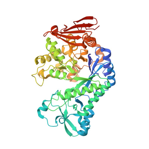
    5M9X, 5MAN

  • PubMed Abstract: 

    Here we present a point mutation-triggered domain shift which switches the acceptor preference of a sucrose phosphorylase from phosphate to a variety of large polyphenolic compounds including resveratrol and quercetin, enabling their efficient glucosylation. The variant possesses a high affinity for aromatic substrates due to newly introduced π-π- and hydrophobic interactions in the altered active site. The domain shift brings about a substantially enlarged and multifunctional active site for polyphenol glucosylation and rare disaccharide production. The crystal structure of the variant with its product resveratrol-3-α-d-glucoside allows the prediction of the substrate scope and regioselectivity of the aromatic compounds' glucosylation sites.


  • Organizational Affiliation

    Department of Organic Chemistry, Universität Würzburg, Am Hubland, 97074 Würzburg, Germany. seibel@chemie.uni-wuerzburg.de.


Macromolecules
Find similar proteins by:  (by identity cutoff)  |  3D Structure
Entity ID: 1
MoleculeChains Sequence LengthOrganismDetailsImage
Sucrose phosphorylaseA [auth B]504Bifidobacterium adolescentisMutation(s): 0 
Gene Names: sucP
EC: 2.4.1.7
UniProt
Find proteins for A0ZZH6 (Bifidobacterium adolescentis (strain ATCC 15703 / DSM 20083 / NCTC 11814 / E194a))
Explore A0ZZH6 
Go to UniProtKB:  A0ZZH6
Entity Groups  
Sequence Clusters30% Identity50% Identity70% Identity90% Identity95% Identity100% Identity
UniProt GroupA0ZZH6
Sequence Annotations
Expand
  • Reference Sequence
Small Molecules
Ligands 1 Unique
IDChains Name / Formula / InChI Key2D Diagram3D Interactions
7KP
Query on 7KP

Download Ideal Coordinates CCD File 
B
(2~{R},3~{S},4~{S},5~{R},6~{R})-2-(hydroxymethyl)-6-[3-[(~{E})-2-(4-hydroxyphenyl)ethenyl]-5-oxidanyl-phenoxy]oxane-3,4 ,5-triol
C20 H22 O8
HSTZMXCBWJGKHG-UABZBGRASA-N
Experimental Data & Validation

Experimental Data

  • Method: X-RAY DIFFRACTION
  • Resolution: 2.35 Å
  • R-Value Free: 0.285 
  • R-Value Work: 0.258 
  • R-Value Observed: 0.260 
  • Space Group: P 43 21 2
Unit Cell:
Length ( Å )Angle ( ˚ )
a = 83.103α = 90
b = 83.103β = 90
c = 157.209γ = 90
Software Package:
Software NamePurpose
PHENIXrefinement
XSCALEdata scaling
PHASERphasing
PDB_EXTRACTdata extraction
PHASERphasing

Structure Validation

View Full Validation Report


Ligand Structure Quality Assessment 


Entry History 

Deposition Data

Revision History  (Full details and data files)

  • Version 1.0: 2017-12-20
    Type: Initial release
  • Version 1.1: 2019-10-16
    Changes: Data collection