5JVP

The neck-linker and alpha 7 helix of Homo sapiens CENP-E

  • Classification: MOTOR PROTEIN
  • Organism(s): Homo sapiens
  • Expression System: Escherichia coli BL21(DE3)
  • Mutation(s): No 

  • Deposited: 2016-05-11 Released: 2016-08-03 
  • Deposition Author(s): Phillips, R.K., Rayment, I.
  • Funding Organization(s): National Institutes of Health/National Institute of General Medical Sciences (NIH/NIGMS), National Science Foundation (NSF, United States)

Experimental Data Snapshot

  • Method: X-RAY DIFFRACTION
  • Resolution: 2.10 Å
  • R-Value Free: 0.274 
  • R-Value Work: 0.237 
  • R-Value Observed: 0.239 

wwPDB Validation   3D Report Full Report


This is version 1.4 of the entry. See complete history


Literature

Family-specific Kinesin Structures Reveal Neck-linker Length Based on Initiation of the Coiled-coil.

Phillips, R.K.Peter, L.G.Gilbert, S.P.Rayment, I.

(2016) J Biol Chem 291: 20372-20386

  • DOI: https://doi.org/10.1074/jbc.M116.737577
  • Primary Citation of Related Structures:  
    5JV3, 5JVM, 5JVP, 5JVR, 5JVS, 5JVU, 5JX1

  • PubMed Abstract: 

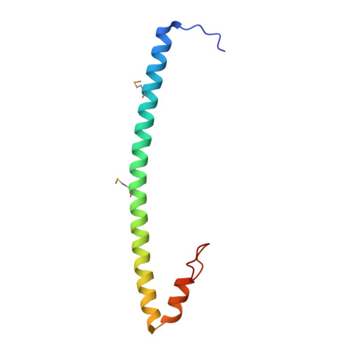
    Kinesin-1, -2, -5, and -7 generate processive hand-over-hand 8-nm steps to transport intracellular cargoes toward the microtubule plus end. This processive motility requires gating mechanisms to coordinate the mechanochemical cycles of the two motor heads to sustain the processive run. A key structural element believed to regulate the degree of processivity is the neck-linker, a short peptide of 12-18 residues, which connects the motor domain to its coiled-coil stalk. Although a shorter neck-linker has been correlated with longer run lengths, the structural data to support this hypothesis have been lacking. To test this hypothesis, seven kinesin structures were determined by x-ray crystallography. Each included the neck-linker motif, followed by helix α7 that constitutes the start of the coiled-coil stalk. In the majority of the structures, the neck-linker length differed from predictions because helix α7, which initiates the coiled-coil, started earlier in the sequence than predicted. A further examination of structures in the Protein Data Bank reveals that there is a great disparity between the predicted and observed starting residues. This suggests that an accurate prediction of the start of a coiled-coil is currently difficult to achieve. These results are significant because they now exclude simple comparisons between members of the kinesin superfamily and add a further layer of complexity when interpreting the results of mutagenesis or protein fusion. They also re-emphasize the need to consider factors beyond the kinesin neck-linker motif when attempting to understand how inter-head communication is tuned to achieve the degree of processivity required for cellular function.


  • Organizational Affiliation

    From the Department of Biochemistry, University of Wisconsin, Madison, Wisconsin 53706 and.


Macromolecules
Find similar proteins by:  (by identity cutoff)  |  3D Structure
Entity ID: 1
MoleculeChains Sequence LengthOrganismDetailsImage
Chimera protein of Centromere-associated protein E and Microtubule-associated protein RP/EB family member 1
A, B, C, D, E
A, B, C, D, E, F
90Homo sapiensMutation(s): 0 
Gene Names: CENPEMAPRE1
UniProt & NIH Common Fund Data Resources
Find proteins for Q02224 (Homo sapiens)
Explore Q02224 
Go to UniProtKB:  Q02224
PHAROS:  Q02224
GTEx:  ENSG00000138778 
Find proteins for Q15691 (Homo sapiens)
Explore Q15691 
Go to UniProtKB:  Q15691
PHAROS:  Q15691
GTEx:  ENSG00000101367 
Entity Groups  
Sequence Clusters30% Identity50% Identity70% Identity90% Identity95% Identity100% Identity
UniProt GroupsQ15691Q02224
Sequence Annotations
Expand
  • Reference Sequence
Small Molecules
Modified Residues  1 Unique
IDChains TypeFormula2D DiagramParent
MSE
Query on MSE
A, B, C, D, E
A, B, C, D, E, F
L-PEPTIDE LINKINGC5 H11 N O2 SeMET
Experimental Data & Validation

Experimental Data

  • Method: X-RAY DIFFRACTION
  • Resolution: 2.10 Å
  • R-Value Free: 0.274 
  • R-Value Work: 0.237 
  • R-Value Observed: 0.239 
  • Space Group: P 32 2 1
Unit Cell:
Length ( Å )Angle ( ˚ )
a = 100.609α = 90
b = 100.609β = 90
c = 107.627γ = 120
Software Package:
Software NamePurpose
PHENIXrefinement
HKL-3000data reduction
HKL-3000data scaling
PHASERphasing

Structure Validation

View Full Validation Report


Entry History & Funding Information

Deposition Data


Funding OrganizationLocationGrant Number
National Institutes of Health/National Institute of General Medical Sciences (NIH/NIGMS)United StatesR37-GM54141
National Science Foundation (NSF, United States)United StatesDGE-0718123

Revision History  (Full details and data files)

  • Version 1.0: 2016-08-03
    Type: Initial release
  • Version 1.1: 2016-08-10
    Changes: Database references
  • Version 1.2: 2016-10-05
    Changes: Database references
  • Version 1.3: 2017-09-27
    Changes: Author supporting evidence, Database references, Derived calculations
  • Version 1.4: 2019-11-27
    Changes: Author supporting evidence