Synthetic, enzyme kinetic, and protein crystallographic studies of C-beta-d-glucopyranosyl pyrroles and imidazoles reveal and explain low nanomolar inhibition of human liver glycogen phosphorylase.
Kantsadi, A.L., Bokor, E., Kun, S., Stravodimos, G.A., Chatzileontiadou, D.S., Leonidas, D.D., Juhasz-Toth, E., Szakacs, A., Batta, G., Docsa, T., Gergely, P., Somsak, L.(2016) Eur J Med Chem 123: 737-745
- PubMed: 27522507
- DOI: https://doi.org/10.1016/j.ejmech.2016.06.049
- Primary Citation of Related Structures:
5JTT, 5JTU, 5LRE - PubMed Abstract:
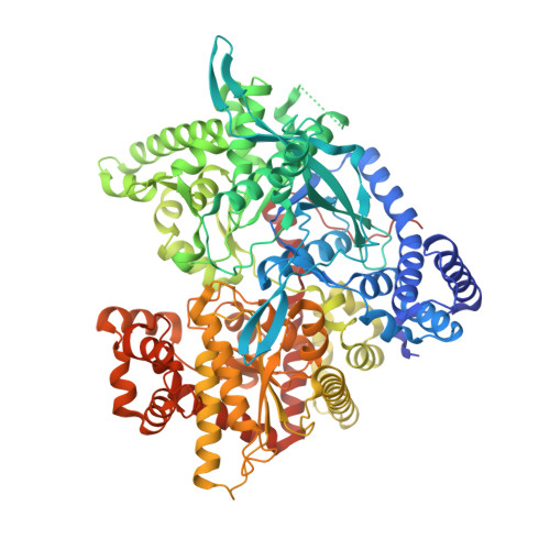
C-β-d-Glucopyranosyl pyrrole derivatives were prepared in the reactions of pyrrole, 2-, and 3-aryl-pyrroles with O-peracetylated β-d-glucopyranosyl trichloroacetimidate, while 2-(β-d-glucopyranosyl) indole was obtained by a cross coupling of O-perbenzylated β-d-glucopyranosyl acetylene with N-tosyl-2-iodoaniline followed by spontaneous ring closure. An improved synthesis of O-perbenzoylated 2-(β-d-glucopyranosyl) imidazoles was achieved by reacting C-glucopyranosyl formimidates with α-aminoketones. The deprotected compounds were assayed with isoforms of glycogen phosphorylase (GP) to show no activity of the pyrroles against rabbit muscle GPb. The imidazoles proved to be the best known glucose derived inhibitors of not only the muscle enzymes (both a and b) but also of the pharmacologically relevant human liver GPa (Ki = 156 and 26 nM for the 4(5)-phenyl and -(2-naphthyl) derivatives, respectively). An X-ray crystallographic study of the rmGPb-imidazole complexes revealed structural features of the strong binding, and also allowed to explain the absence of inhibition for the pyrrole derivatives.
Organizational Affiliation:
Department of Biochemistry and Biotechnology, University of Thessaly, 26 Ploutonos Str. 41221, Larissa, Greece.