5J3H

Human insulin receptor domains L1-CR in complex with peptide S519C16 and 83-7 Fv


Experimental Data Snapshot

  • Method: X-RAY DIFFRACTION
  • Resolution: 3.27 Å
  • R-Value Free: 0.248 
  • R-Value Work: 0.226 
  • R-Value Observed: 0.227 

wwPDB Validation   3D Report Full Report


Ligand Structure Quality Assessment 


This is version 2.1 of the entry. See complete history


Literature

Insulin Mimetic Peptide Disrupts the Primary Binding Site of the Insulin Receptor.

Lawrence, C.F.Margetts, M.B.Menting, J.G.Smith, N.A.Smith, B.J.Ward, C.W.Lawrence, M.C.

(2016) J Biol Chem 291: 15473-15481

  • DOI: https://doi.org/10.1074/jbc.M116.732180
  • Primary Citation of Related Structures:  
    5J3H

  • PubMed Abstract: 

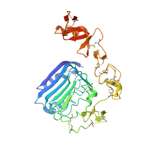
    Sets of synthetic peptides that interact with the insulin receptor ectodomain have been discovered by phage display and reported in the literature. These peptides were grouped into three classes termed Site 1, Site 2, and Site 3 based on their mutual competition of binding to the receptor. Further refinement has yielded, in particular, a 36-residue Site 2-Site 1 fusion peptide, S519, that binds the insulin receptor with subnanomolar affinity and exhibits agonist activity in both lipogenesis and glucose uptake assays. Here, we report three-dimensional crystallographic detail of the interaction of the C-terminal, 16-residue Site 1 component (S519C16) of S519 with the first leucine-rich repeat domain (L1) of the insulin receptor. Our structure shows that S519C16 binds to the same site on the L1 surface as that occupied by a critical component of the primary binding site, namely the helical C-terminal segment of the insulin receptor α-chain (termed αCT). In particular, the two phenylalanine residues within the FYXWF motif of S519C16 are seen to engage the insulin receptor L1 domain surface in a fashion almost identical to the respective αCT residues Phe(701) and Phe(705) The structure provides a platform for the further development of peptidic and/or small molecule agents directed toward the insulin receptor and/or the type 1 insulin-like growth factor receptor.


  • Organizational Affiliation

    From the Walter and Eliza Hall Institute of Medical Research, 1G Royal Parade, Parkville, Victoria 3052, Australia.


Macromolecules

Find similar proteins by:  Sequence   |   3D Structure  

Entity ID: 1
MoleculeChains Sequence LengthOrganismDetailsImage
Peptide S519C16A [auth B]16Endothia gyrosaMutation(s): 0 
Entity Groups  
Sequence Clusters30% Identity50% Identity70% Identity90% Identity95% Identity100% Identity
Sequence Annotations
Expand
  • Reference Sequence
Find similar proteins by:  (by identity cutoff)  |  3D Structure
Entity ID: 2
MoleculeChains Sequence LengthOrganismDetailsImage
mAb 83-7 Fab heavy chainB [auth C]123Mus musculusMutation(s): 0 
Entity Groups  
Sequence Clusters30% Identity50% Identity70% Identity90% Identity95% Identity100% Identity
Sequence Annotations
Expand
  • Reference Sequence
Find similar proteins by:  (by identity cutoff)  |  3D Structure
Entity ID: 3
MoleculeChains Sequence LengthOrganismDetailsImage
mAb 83-7 Fab light chainC [auth D]118Mus musculusMutation(s): 0 
Entity Groups  
Sequence Clusters30% Identity50% Identity70% Identity90% Identity95% Identity100% Identity
Sequence Annotations
Expand
  • Reference Sequence
Find similar proteins by:  (by identity cutoff)  |  3D Structure
Entity ID: 4
MoleculeChains Sequence LengthOrganismDetailsImage
Insulin receptorD [auth E]317Homo sapiensMutation(s): 0 
Gene Names: INSR
EC: 2.7.10.1
UniProt & NIH Common Fund Data Resources
Find proteins for P06213 (Homo sapiens)
Explore P06213 
Go to UniProtKB:  P06213
PHAROS:  P06213
GTEx:  ENSG00000171105 
Entity Groups  
Sequence Clusters30% Identity50% Identity70% Identity90% Identity95% Identity100% Identity
UniProt GroupP06213
Sequence Annotations
Expand
  • Reference Sequence
Oligosaccharides

Help

Entity ID: 5
MoleculeChains Length2D Diagram Glycosylation3D Interactions
2-acetamido-2-deoxy-beta-D-glucopyranose-(1-4)-[alpha-L-fucopyranose-(1-6)]2-acetamido-2-deoxy-beta-D-glucopyranoseE [auth A]3N-Glycosylation
Glycosylation Resources
GlyTouCan:  G21290RB
GlyCosmos:  G21290RB
GlyGen:  G21290RB
Entity ID: 6
MoleculeChains Length2D Diagram Glycosylation3D Interactions
2-acetamido-2-deoxy-beta-D-glucopyranose-(1-4)-2-acetamido-2-deoxy-beta-D-glucopyranose
F
2N-Glycosylation
Glycosylation Resources
GlyTouCan:  G42666HT
GlyCosmos:  G42666HT
GlyGen:  G42666HT
Entity ID: 7
MoleculeChains Length2D Diagram Glycosylation3D Interactions
2-acetamido-2-deoxy-beta-D-glucopyranose-(1-2)-alpha-D-mannopyranose-(1-3)-[alpha-D-mannopyranose-(1-6)]beta-D-mannopyranose-(1-4)-2-acetamido-2-deoxy-beta-D-glucopyranose-(1-4)-[alpha-L-fucopyranose-(1-6)]2-acetamido-2-deoxy-beta-D-glucopyranose
G
7N-Glycosylation
Glycosylation Resources
GlyTouCan:  G07483YN
GlyCosmos:  G07483YN
GlyGen:  G07483YN
Small Molecules
Ligands 1 Unique
IDChains Name / Formula / InChI Key2D Diagram3D Interactions
NAG
Query on NAG

Download Ideal Coordinates CCD File 
H [auth E],
I [auth E]
2-acetamido-2-deoxy-beta-D-glucopyranose
C8 H15 N O6
OVRNDRQMDRJTHS-FMDGEEDCSA-N
Experimental Data & Validation

Experimental Data

  • Method: X-RAY DIFFRACTION
  • Resolution: 3.27 Å
  • R-Value Free: 0.248 
  • R-Value Work: 0.226 
  • R-Value Observed: 0.227 
  • Space Group: P 41 21 2
Unit Cell:
Length ( Å )Angle ( ˚ )
a = 176.776α = 90
b = 176.776β = 90
c = 86.186γ = 90
Software Package:
Software NamePurpose
BUSTERrefinement
XDSdata reduction
XSCALEdata scaling
PHASERphasing

Structure Validation

View Full Validation Report


Ligand Structure Quality Assessment 


Entry History & Funding Information

Deposition Data


Funding OrganizationLocationGrant Number
National Health and Medical Research Council (NHMRC, Australia)Australia1005896
National Health and Medical Research Council (NHMRC, Australia)Australia1058233
National Health and Medical Research Council (NHMRC, Australia)Australia361646
Victorian State GovernmentAustraliaOperational Infrastructure Support Grant

Revision History  (Full details and data files)

  • Version 1.0: 2016-06-15
    Type: Initial release
  • Version 1.1: 2016-06-22
    Changes: Database references
  • Version 1.2: 2016-08-10
    Changes: Database references
  • Version 1.3: 2017-09-13
    Changes: Author supporting evidence, Data collection, Database references, Derived calculations
  • Version 1.4: 2020-01-08
    Changes: Author supporting evidence, Data collection
  • Version 2.0: 2020-07-29
    Type: Remediation
    Reason: Carbohydrate remediation
    Changes: Atomic model, Data collection, Derived calculations, Structure summary
  • Version 2.1: 2023-09-27
    Changes: Data collection, Database references, Derived calculations, Refinement description, Structure summary