5IR3

Crystal structure of the recombinant highest fibrillogenic natural mutant (obtained from patient AR) derived from lambda 6 light chain variable domain


Experimental Data Snapshot

  • Method: X-RAY DIFFRACTION
  • Resolution: 1.70 Å
  • R-Value Free: 0.207 
  • R-Value Work: 0.174 
  • R-Value Observed: 0.176 

wwPDB Validation   3D Report Full Report


This is version 1.3 of the entry. See complete history


Literature

Stabilizing an amyloidogenic lambda 6 light chain variable domain.

Luna-Martinez, O.D.Hernandez-Santoyo, A.Villalba-Velazquez, M.I.Sanchez-Alcala, R.Fernandez-Velasco, D.A.Becerril, B.

(2017) FEBS J 284: 3702-3717

  • DOI: https://doi.org/10.1111/febs.14265
  • Primary Citation of Related Structures:  
    5IR3

  • PubMed Abstract: 

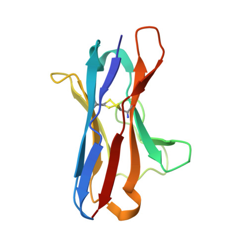
    Light chain amyloidosis is a lethal disease where vital organs are damaged by the fibrillar aggregation of monoclonal light chains. λ6a is an immunoglobulin light chain encoded by the germ-line gene segment implicated in this disease. AR is a patient-derived germ-line variant with a markedly low thermodynamic stability and prone to form fibrils in vitro in less than an hour. Here, we sought to stabilize this domain by mutating some residues back to the germ-line sequence, and the most stabilizing mutations were the single-mutant AR-F21I and the double-mutant AR-F21/IV104L, both located in the hydrophobic core. While mutation Arg25Gly in 6aJL2 destabilized the domain, mutating Gly25 back to arginine in AR did not contribute to stabilization as expected. Crystallographic structures of AR and 6a-R25G were generated to explain this discrepancy. Finally, 6a-R25G crystals revealed an octameric assembly which was emulated into 6aJL2 and AR crystals by replicating their structural parameters and suggesting a common assembly pattern.


  • Organizational Affiliation

    Departamento de Medicina Molecular y Bioprocesos, Instituto de Biotecnología, Universidad Nacional Autónoma de México, Cuernavaca, Mexico.


Macromolecules
Find similar proteins by:  (by identity cutoff)  |  3D Structure
Entity ID: 1
MoleculeChains Sequence LengthOrganismDetailsImage
Ig lambda chain V-VI region AR112Homo sapiensMutation(s): 0 
UniProt & NIH Common Fund Data Resources
Find proteins for P01721 (Homo sapiens)
Explore P01721 
Go to UniProtKB:  P01721
PHAROS:  P01721
GTEx:  ENSG00000211640 
Entity Groups  
Sequence Clusters30% Identity50% Identity70% Identity90% Identity95% Identity100% Identity
UniProt GroupP01721
Sequence Annotations
Expand
  • Reference Sequence
Small Molecules
Ligands 1 Unique
IDChains Name / Formula / InChI Key2D Diagram3D Interactions
ACT
Query on ACT

Download Ideal Coordinates CCD File 
B [auth A]ACETATE ION
C2 H3 O2
QTBSBXVTEAMEQO-UHFFFAOYSA-M
Experimental Data & Validation

Experimental Data

  • Method: X-RAY DIFFRACTION
  • Resolution: 1.70 Å
  • R-Value Free: 0.207 
  • R-Value Work: 0.174 
  • R-Value Observed: 0.176 
  • Space Group: I 41 2 2
Unit Cell:
Length ( Å )Angle ( ˚ )
a = 71.121α = 90
b = 71.121β = 90
c = 96.453γ = 90
Software Package:
Software NamePurpose
PHENIXrefinement
XDSdata reduction
SCALAdata scaling
PHASERphasing

Structure Validation

View Full Validation Report


Entry History 

Deposition Data

Revision History  (Full details and data files)

  • Version 1.0: 2017-06-14
    Type: Initial release
  • Version 1.1: 2017-09-27
    Changes: Database references
  • Version 1.2: 2017-11-22
    Changes: Database references
  • Version 1.3: 2023-09-27
    Changes: Data collection, Database references, Refinement description