5IF6

Structure of a computationally designed 17-OHP binder


Experimental Data Snapshot

  • Method: X-RAY DIFFRACTION
  • Resolution: 2.50 Å
  • R-Value Free: 0.244 
  • R-Value Work: 0.213 
  • R-Value Observed: 0.215 

wwPDB Validation   3D Report Full Report


Ligand Structure Quality Assessment 


This is version 1.4 of the entry. See complete history


Literature

Sampling and energy evaluation challenges in ligand binding protein design.

Dou, J.Doyle, L.Jr Greisen, P.Schena, A.Park, H.Johnsson, K.Stoddard, B.L.Baker, D.

(2017) Protein Sci 26: 2426-2437

  • DOI: https://doi.org/10.1002/pro.3317
  • Primary Citation of Related Structures:  
    5IER, 5IF6

  • PubMed Abstract: 

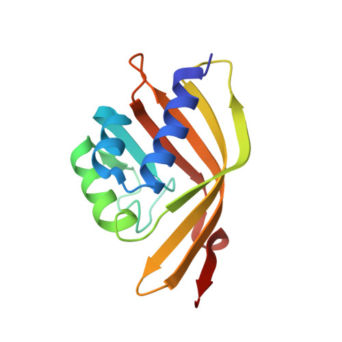
    The steroid hormone 17α-hydroxylprogesterone (17-OHP) is a biomarker for congenital adrenal hyperplasia and hence there is considerable interest in development of sensors for this compound. We used computational protein design to generate protein models with binding sites for 17-OHP containing an extended, nonpolar, shape-complementary binding pocket for the four-ring core of the compound, and hydrogen bonding residues at the base of the pocket to interact with carbonyl and hydroxyl groups at the more polar end of the ligand. Eight of 16 designed proteins experimentally tested bind 17-OHP with micromolar affinity. A co-crystal structure of one of the designs revealed that 17-OHP is rotated 180° around a pseudo-two-fold axis in the compound and displays multiple binding modes within the pocket, while still interacting with all of the designed residues in the engineered site. Subsequent rounds of mutagenesis and binding selection improved the ligand affinity to nanomolar range, while appearing to constrain the ligand to a single bound conformation that maintains the same "flipped" orientation relative to the original design. We trace the discrepancy in the design calculations to two sources: first, a failure to model subtle backbone changes which alter the distribution of sidechain rotameric states and second, an underestimation of the energetic cost of desolvating the carbonyl and hydroxyl groups of the ligand. The difference between design model and crystal structure thus arises from both sampling limitations and energy function inaccuracies that are exacerbated by the near two-fold symmetry of the molecule.


  • Organizational Affiliation

    Department of Bioengineering, University of Washington, Seattle, Washington.


Macromolecules
Find similar proteins by:  (by identity cutoff)  |  3D Structure
Entity ID: 1
MoleculeChains Sequence LengthOrganismDetailsImage
OHP9_1c
A, B, C, D, E
A, B, C, D, E, F
134synthetic constructMutation(s): 0 
Entity Groups  
Sequence Clusters30% Identity50% Identity70% Identity90% Identity95% Identity100% Identity
Sequence Annotations
Expand
  • Reference Sequence
Small Molecules
Ligands 2 Unique
IDChains Name / Formula / InChI Key2D Diagram3D Interactions
3QZ
Query on 3QZ

Download Ideal Coordinates CCD File 
G [auth A]
I [auth B]
K [auth C]
L [auth D]
N [auth E]
G [auth A],
I [auth B],
K [auth C],
L [auth D],
N [auth E],
O [auth F]
(9beta)-17-hydroxypregn-4-ene-3,20-dione
C21 H30 O3
DBPWSSGDRRHUNT-CEGNMAFCSA-N
NA
Query on NA

Download Ideal Coordinates CCD File 
H [auth A],
J [auth B],
M [auth D]
SODIUM ION
Na
FKNQFGJONOIPTF-UHFFFAOYSA-N
Experimental Data & Validation

Experimental Data

  • Method: X-RAY DIFFRACTION
  • Resolution: 2.50 Å
  • R-Value Free: 0.244 
  • R-Value Work: 0.213 
  • R-Value Observed: 0.215 
  • Space Group: P 21 21 21
Unit Cell:
Length ( Å )Angle ( ˚ )
a = 80.02α = 90
b = 100.787β = 90
c = 118.154γ = 90
Software Package:
Software NamePurpose
PHENIXrefinement
HKL-2000data scaling
PHASERphasing
PDB_EXTRACTdata extraction
HKL-2000data reduction

Structure Validation

View Full Validation Report


Ligand Structure Quality Assessment 


Entry History & Funding Information

Deposition Data


Funding OrganizationLocationGrant Number
National Institutes of Health/National Institute of General Medical Sciences (NIH/NIGMS)United StatesGM115545

Revision History  (Full details and data files)

  • Version 1.0: 2017-03-01
    Type: Initial release
  • Version 1.1: 2017-09-27
    Changes: Author supporting evidence
  • Version 1.2: 2018-02-07
    Changes: Database references
  • Version 1.3: 2019-12-25
    Changes: Author supporting evidence
  • Version 1.4: 2023-09-27
    Changes: Data collection, Database references, Refinement description