Novel insights into the interaction of UBA5 with UFM1 via a UFM1-interacting sequence.
Padala, P., Oweis, W., Mashahreh, B., Soudah, N., Cohen-Kfir, E., Todd, E.A., Berndsen, C.E., Wiener, R.(2017) Sci Rep 7: 508-508
- PubMed: 28360427
- DOI: https://doi.org/10.1038/s41598-017-00610-0
- Primary Citation of Related Structures:
5IA7, 5IA8 - PubMed Abstract:
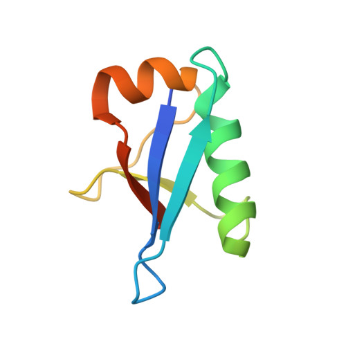
The modification of proteins by ubiquitin-fold modifier 1 (UFM1) is implicated in many human diseases. Prior to conjugation, UFM1 undergoes activation by its cognate activating enzyme, UBA5. UBA5 is a non-canonical E1 activating enzyme that possesses an adenylation domain but lacks a distinct cysteine domain. Binding of UBA5 to UFM1 is mediated via an amino acid sequence, known as the UFM1-interacting sequence (UIS), located outside the adenylation domain that is required for UFM1 activation. However, the precise boundaries of the UIS are yet not clear and are still under debate. Here we revisit the interaction of UFM1 with UBA5 by determining the crystal structure of UFM1 fused to 13 amino acids of human UBA5. Using binding and activity assays, we found that His 336 of UBA5, previously not reported to be part of the UIS, occupies a negatively charged pocket on UFM1's surface. This His is involved in UFM1 binding and if mutated perturbs activation of UFM1. Surprisingly, we also found that the interaction between two UFM1 molecules mimics how the UIS binds UFM1. Specifically, UFM1 His 70 resembles UBA5 His336 and enters a negatively charged pocked on the other UFM1 molecule. Our results refine our understanding of UFM1-UBA5 binding.
Organizational Affiliation:
Department of Biochemistry and Molecular Biology, the Institute for Medical Research Israel-Canada, Hebrew University-Hadassah Medical School, Jerusalem, 91120, Israel.