5GO9

Cryo-EM structure of RyR2 in closed state

  • Classification: TRANSPORT PROTEIN
  • Organism(s): Sus scrofa
  • Mutation(s): No 

  • Deposited: 2016-07-26 Released: 2016-10-05 
  • Deposition Author(s): Peng, W., Wu, J.P., Yan, N.
  • Funding Organization(s): Ministry of Science and Technology of China, National Natural Science Foundation of China

Experimental Data Snapshot

  • Method: ELECTRON MICROSCOPY
  • Resolution: 4.40 Å
  • Aggregation State: PARTICLE 
  • Reconstruction Method: SINGLE PARTICLE 

wwPDB Validation   3D Report Full Report


This is version 1.3 of the entry. See complete history


Literature

Structural basis for the gating mechanism of the type 2 ryanodine receptor RyR2

Peng, W.Shen, H.Wu, J.P.Guo, W.Pan, X.Wang, R.Chen, S.R.W.Yan, N.

(2016) Science 354

  • DOI: https://doi.org/10.1126/science.aah5324
  • Primary Citation of Related Structures:  
    5GO9, 5GOA

  • PubMed Abstract: 

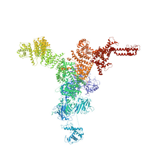
    RyR2 is a high-conductance intracellular calcium (Ca 2+ ) channel that controls the release of Ca 2+ from the sarco(endo)plasmic reticulum of a variety of cells. Here, we report the structures of RyR2 from porcine heart in both the open and closed states at near-atomic resolutions determined using single-particle electron cryomicroscopy. Structural comparison reveals a breathing motion of the overall cytoplasmic region resulted from the interdomain movements of amino-terminal domains (NTDs), Helical domains, and Handle domains, whereas almost no intradomain shifts are observed in these armadillo repeats-containing domains. Outward rotations of the Central domains, which integrate the conformational changes of the cytoplasmic region, lead to the dilation of the cytoplasmic gate through coupled motions. Our structural and mutational characterizations provide important insights into the gating and disease mechanism of RyRs.


  • Organizational Affiliation

    State Key Laboratory of Membrane Biology, Tsinghua University, Beijing 100084, China.


Macromolecules
Find similar proteins by:  (by identity cutoff)  |  3D Structure
Entity ID: 1
MoleculeChains Sequence LengthOrganismDetailsImage
RyR2
A, B, C, D
4,968Sus scrofaMutation(s): 0 
Entity Groups  
Sequence Clusters30% Identity50% Identity70% Identity90% Identity95% Identity100% Identity
Sequence Annotations
Expand
  • Reference Sequence
Experimental Data & Validation

Experimental Data

  • Method: ELECTRON MICROSCOPY
  • Resolution: 4.40 Å
  • Aggregation State: PARTICLE 
  • Reconstruction Method: SINGLE PARTICLE 
EM Software:
TaskSoftware PackageVersion
MODEL REFINEMENTREFMAC5.8
RECONSTRUCTIONRELION1.4

Structure Validation

View Full Validation Report


Entry History & Funding Information

Deposition Data


Funding OrganizationLocationGrant Number
Ministry of Science and Technology of ChinaChina2015CB9101012014, 2016YFA0500402, ZX09507003006
National Natural Science Foundation of ChinaChinaproject 31321062, 31021002, 31611130036

Revision History  (Full details and data files)

  • Version 1.0: 2016-10-05
    Type: Initial release
  • Version 1.1: 2017-02-08
    Changes: Database references
  • Version 1.2: 2019-11-06
    Changes: Data collection, Other
  • Version 1.3: 2024-03-27
    Changes: Data collection, Database references