5GMX

Crystal structure of a family VIII carboxylesterase


Experimental Data Snapshot

  • Method: X-RAY DIFFRACTION
  • Resolution: 2.00 Å
  • R-Value Free: 0.222 
  • R-Value Work: 0.184 
  • R-Value Observed: 0.186 

wwPDB Validation   3D Report Full Report


Ligand Structure Quality Assessment 


This is version 1.1 of the entry. See complete history


Literature

Crystal structure of EstSRT1, a family VIII carboxylesterase displaying hydrolytic activity toward oxyimino cephalosporins

Cha, S.S.An, Y.J.

(2016) Biochem Biophys Res Commun 478: 818-824

  • DOI: https://doi.org/10.1016/j.bbrc.2016.08.031
  • Primary Citation of Related Structures:  
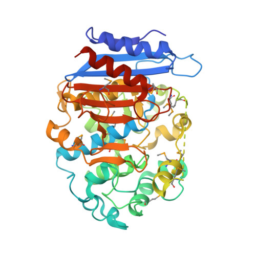
    5GMX

  • PubMed Abstract: 

    EstSRT1 is a family VIII carboxylesterase that hydrolyzes oxyimino third- and fourth-generation cephalosporins, first-generation cephalosporins and ester substrates. According to the crystal structure of EstSRT1 (2.0-Å resolution), this protein contains a large α/β domain and a small α-helical domain and harbors three catalytic residues (Ser71, Lys74, and Tyr160) in the cavity at the domain interface, similarly to other family VIII carboxylesterases. Comparison of the structures of EstSRT1 and EstU1, a family VIII carboxylesterase with no hydrolytic activity toward bulky oxyimino cephalosporins, revealed that EstSRT1 has a smaller active site, despite its extended substrate range. The B-factors of the active site segments that could potentially contact with the oxyimino groups and the R2 side chains of oxyimino cephalosporins are higher in EstSRT1 than in EstU1, thus suggesting the role of the active site's structural flexibility in the extension of EstSRT1's substrate spectrum.


  • Organizational Affiliation

    Department of Chemistry & Nano Science, Ewha Womans University, Seoul, 03760, Republic of Korea. Electronic address: chajung@ewha.ac.kr.


Macromolecules
Find similar proteins by:  (by identity cutoff)  |  3D Structure
Entity ID: 1
MoleculeChains Sequence LengthOrganismDetailsImage
Carboxylesterase
A, B
398uncultured bacteriumMutation(s): 0 
EC: 3.1.1.1
UniProt
Find proteins for A0A0G2RKR9 (uncultured bacterium)
Explore A0A0G2RKR9 
Go to UniProtKB:  A0A0G2RKR9
Entity Groups  
Sequence Clusters30% Identity50% Identity70% Identity90% Identity95% Identity100% Identity
UniProt GroupA0A0G2RKR9
Sequence Annotations
Expand
  • Reference Sequence
Small Molecules
Modified Residues  1 Unique
IDChains TypeFormula2D DiagramParent
MSE
Query on MSE
A, B
L-PEPTIDE LINKINGC5 H11 N O2 SeMET
Experimental Data & Validation

Experimental Data

  • Method: X-RAY DIFFRACTION
  • Resolution: 2.00 Å
  • R-Value Free: 0.222 
  • R-Value Work: 0.184 
  • R-Value Observed: 0.186 
  • Space Group: P 21 21 21
Unit Cell:
Length ( Å )Angle ( ˚ )
a = 80.128α = 90
b = 95.149β = 90
c = 99.943γ = 90
Software Package:
Software NamePurpose
PHENIXrefinement
HKL-2000data reduction
HKL-2000data scaling
SOLVEphasing

Structure Validation

View Full Validation Report


Ligand Structure Quality Assessment 


Entry History 

Deposition Data

Revision History  (Full details and data files)

  • Version 1.0: 2017-07-26
    Type: Initial release
  • Version 1.1: 2019-02-06
    Changes: Data collection, Database references