5GG4

Crystal structure of USP7 with RNF169 peptide


Experimental Data Snapshot

  • Method: X-RAY DIFFRACTION
  • Resolution: 3.11 Å
  • R-Value Free: 0.268 
  • R-Value Work: 0.226 
  • R-Value Observed: 0.228 

wwPDB Validation   3D Report Full Report


This is version 1.3 of the entry. See complete history


Literature

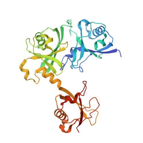
Dual-utility NLS drives RNF169-dependent DNA damage responses.

An, L.Jiang, Y.Ng, H.H.Man, E.P.Chen, J.Khoo, U.S.Gong, Q.Huen, M.S.

(2017) Proc Natl Acad Sci U S A 114: E2872-E2881

  • DOI: https://doi.org/10.1073/pnas.1616602114
  • Primary Citation of Related Structures:  
    5GG4

  • PubMed Abstract: 

    Loading of p53-binding protein 1 (53BP1) and receptor-associated protein 80 (RAP80) at DNA double-strand breaks (DSBs) drives cell cycle checkpoint activation but is counterproductive to high-fidelity DNA repair. ring finger protein 169 (RNF169) maintains the balance by limiting the deposition of DNA damage mediator proteins at the damaged chromatin. We report here that this attribute is accomplished, in part, by a predicted nuclear localization signal (NLS) that not only shuttles RNF169 into the nucleus but also promotes its stability by mediating a direct interaction with the ubiquitin-specific protease USP7. Guided by the crystal structure of USP7 in complex with the RNF169 NLS, we uncoupled USP7 binding from its nuclear import function and showed that perturbing the USP7-RNF169 complex destabilized RNF169, compromised high-fidelity DSB repair, and hypersensitized cells to poly (ADP-ribose) polymerase inhibition. Finally, expression of USP7 and RNF169 positively correlated in breast cancer specimens. Collectively, our findings uncover an NLS-mediated bipartite mechanism that supports the nuclear function of a DSB response protein.


  • Organizational Affiliation

    School of Biomedical Sciences, Li Ka Shing Faculty of Medicine, The University of Hong Kong, Hong Kong Special Administrative Region.


Macromolecules
Find similar proteins by:  (by identity cutoff)  |  3D Structure
Entity ID: 1
MoleculeChains Sequence LengthOrganismDetailsImage
Ubiquitin carboxyl-terminal hydrolase 7
A, B, C, D
334Homo sapiensMutation(s): 0 
Gene Names: USP7HAUSP
EC: 3.4.19.12
UniProt & NIH Common Fund Data Resources
Find proteins for Q93009 (Homo sapiens)
Explore Q93009 
Go to UniProtKB:  Q93009
PHAROS:  Q93009
GTEx:  ENSG00000187555 
Entity Groups  
Sequence Clusters30% Identity50% Identity70% Identity90% Identity95% Identity100% Identity
UniProt GroupQ93009
Sequence Annotations
Expand
  • Reference Sequence

Find similar proteins by:  Sequence   |   3D Structure  

Entity ID: 2
MoleculeChains Sequence LengthOrganismDetailsImage
Peptide from E3 ubiquitin-protein ligase RNF169
E, F, G, H
13Homo sapiensMutation(s): 0 
EC: 6.3.2
UniProt & NIH Common Fund Data Resources
Find proteins for Q8NCN4 (Homo sapiens)
Explore Q8NCN4 
Go to UniProtKB:  Q8NCN4
PHAROS:  Q8NCN4
GTEx:  ENSG00000166439 
Entity Groups  
Sequence Clusters30% Identity50% Identity70% Identity90% Identity95% Identity100% Identity
UniProt GroupQ8NCN4
Sequence Annotations
Expand
  • Reference Sequence
Experimental Data & Validation

Experimental Data

  • Method: X-RAY DIFFRACTION
  • Resolution: 3.11 Å
  • R-Value Free: 0.268 
  • R-Value Work: 0.226 
  • R-Value Observed: 0.228 
  • Space Group: C 1 2 1
Unit Cell:
Length ( Å )Angle ( ˚ )
a = 162.195α = 90
b = 99.965β = 95.54
c = 124.299γ = 90
Software Package:
Software NamePurpose
HKL-2000data scaling
REFMACrefinement
PDB_EXTRACTdata extraction
HKL-2000data reduction
PHENIXphasing

Structure Validation

View Full Validation Report


Entry History 

Deposition Data

Revision History  (Full details and data files)

  • Version 1.0: 2017-03-22
    Type: Initial release
  • Version 1.1: 2017-04-19
    Changes: Database references
  • Version 1.2: 2017-10-04
    Changes: Data collection
  • Version 1.3: 2023-11-08
    Changes: Data collection, Database references, Refinement description