5FOI

Crystal structure of mycinamicin VIII C21 methyl hydroxylase MycCI from Micromonospora griseorubida bound to mycinamicin VIII


Experimental Data Snapshot

  • Method: X-RAY DIFFRACTION
  • Resolution: 2.21 Å
  • R-Value Free: 0.252 
  • R-Value Work: 0.182 
  • R-Value Observed: 0.186 

wwPDB Validation   3D Report Full Report


Ligand Structure Quality Assessment 


This is version 1.2 of the entry. See complete history


Literature

Biochemical and Structural Characterization of Mycci, a Versatile P450 Biocatalyst from the Mycinamicin Biosynthetic Pathway.

Demars, M.D.Sheng, F.Park, S.R.Lowell, A.N.Podust, L.M.Sherman, D.H.

(2016) ACS Chem Biol 11: 2642

  • DOI: https://doi.org/10.1021/acschembio.6b00479
  • Primary Citation of Related Structures:  
    5FOI

  • PubMed Abstract: 

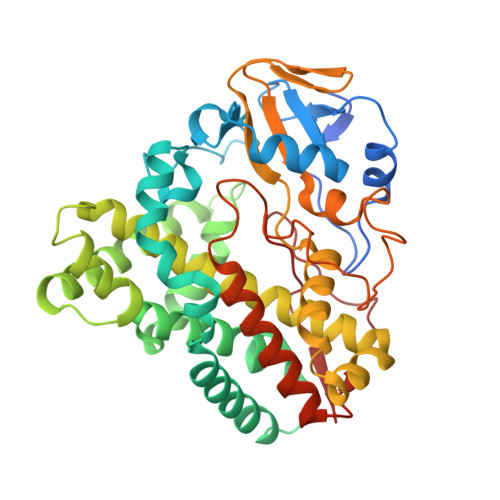
    Cytochrome P450 monooxygenases (P450s) are some of nature's most ubiquitous and versatile enzymes for performing oxidative metabolic transformations. Their unmatched ability to selectively functionalize inert C-H bonds has led to their increasing employment in academic and industrial settings for the production of fine and commodity chemicals. Many of the most interesting and potentially biocatalytically useful P450s come from microorganisms, where they catalyze key tailoring reactions in natural product biosynthetic pathways. While most of these enzymes act on structurally complex pathway intermediates with high selectivity, they often exhibit narrow substrate scope, thus limiting their broader application. In the present study, we investigated the reactivity of the P450 MycCI from the mycinamicin biosynthetic pathway toward a variety of macrocyclic compounds and discovered that the enzyme exhibits appreciable activity on several 16-membered ring macrolactones independent of their glycosylation state. These results were corroborated by performing equilibrium substrate binding experiments, steady-state kinetics studies, and X-ray crystallographic analysis of MycCI bound to its native substrate mycinamicin VIII. We also characterized TylHI, a homologous P450 from the tylosin pathway, and showed that its substrate scope is severely restricted compared to MycCI. Thus, the ability of the latter to hydroxylate both macrocyclic aglycones and macrolides sets it apart from related biosynthetic P450s and highlights its potential for developing novel P450 biocatalysts with broad substrate scope and high regioselectivity.


  • Organizational Affiliation

    Life Sciences Institute, University of Michigan , Ann Arbor, Michigan 48109, United States.


Macromolecules
Find similar proteins by:  (by identity cutoff)  |  3D Structure
Entity ID: 1
MoleculeChains Sequence LengthOrganismDetailsImage
MYCINAMICIN VIII C21 METHYL HYDROXYLASE
A, B
408Micromonospora griseorubidaMutation(s): 0 
EC: 1.14
UniProt
Find proteins for Q83WF5 (Micromonospora griseorubida)
Explore Q83WF5 
Go to UniProtKB:  Q83WF5
Entity Groups  
Sequence Clusters30% Identity50% Identity70% Identity90% Identity95% Identity100% Identity
UniProt GroupQ83WF5
Sequence Annotations
Expand
  • Reference Sequence
Small Molecules
Ligands 4 Unique
IDChains Name / Formula / InChI Key2D Diagram3D Interactions
HEM
Query on HEM

Download Ideal Coordinates CCD File 
C [auth A],
J [auth B]
PROTOPORPHYRIN IX CONTAINING FE
C34 H32 Fe N4 O4
KABFMIBPWCXCRK-RGGAHWMASA-L
MY8
Query on MY8

Download Ideal Coordinates CCD File 
D [auth A],
K [auth B]
Mycinamicin VIII
C29 H49 N O6
ZWHLYXQJICTUSD-JUFBBUGVSA-N
SPD
Query on SPD

Download Ideal Coordinates CCD File 
G [auth A],
H [auth A],
I [auth A]
SPERMIDINE
C7 H19 N3
ATHGHQPFGPMSJY-UHFFFAOYSA-N
GOL
Query on GOL

Download Ideal Coordinates CCD File 
E [auth A],
F [auth A]
GLYCEROL
C3 H8 O3
PEDCQBHIVMGVHV-UHFFFAOYSA-N
Binding Affinity Annotations 
IDSourceBinding Affinity
MY8 Binding MOAD:  5FOI Kd: 1 (nM) from 1 assay(s)
Experimental Data & Validation

Experimental Data

  • Method: X-RAY DIFFRACTION
  • Resolution: 2.21 Å
  • R-Value Free: 0.252 
  • R-Value Work: 0.182 
  • R-Value Observed: 0.186 
  • Space Group: P 1
Unit Cell:
Length ( Å )Angle ( ˚ )
a = 54.781α = 83.2
b = 59.516β = 72.15
c = 74.623γ = 62.61
Software Package:
Software NamePurpose
REFMACrefinement
MOSFLMdata reduction
SCALAdata scaling
MrBUMPphasing

Structure Validation

View Full Validation Report


Ligand Structure Quality Assessment 


Entry History 

Deposition Data

Revision History  (Full details and data files)

  • Version 1.0: 2016-07-27
    Type: Initial release
  • Version 1.1: 2016-10-05
    Changes: Database references
  • Version 1.2: 2024-01-10
    Changes: Data collection, Database references, Derived calculations, Other, Refinement description