5EJK

Crystal structure of the Rous sarcoma virus intasome


Experimental Data Snapshot

  • Method: X-RAY DIFFRACTION
  • Resolution: 3.80 Å
  • R-Value Free: 0.294 
  • R-Value Work: 0.254 
  • R-Value Observed: 0.256 

wwPDB Validation   3D Report Full Report


This is version 1.2 of the entry. See complete history


Literature

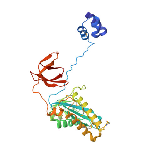
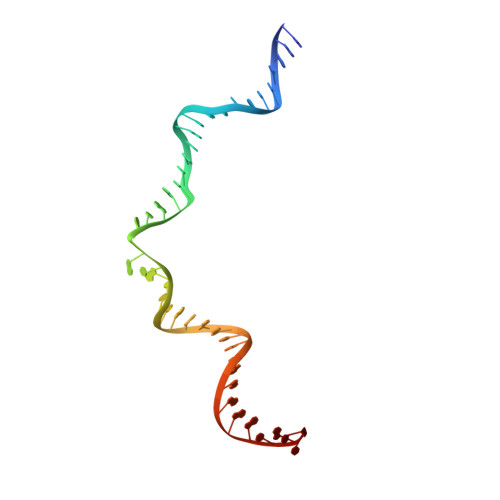
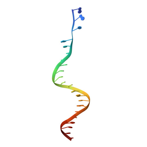
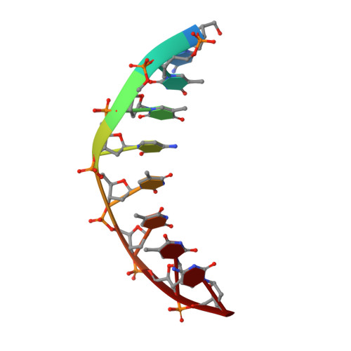
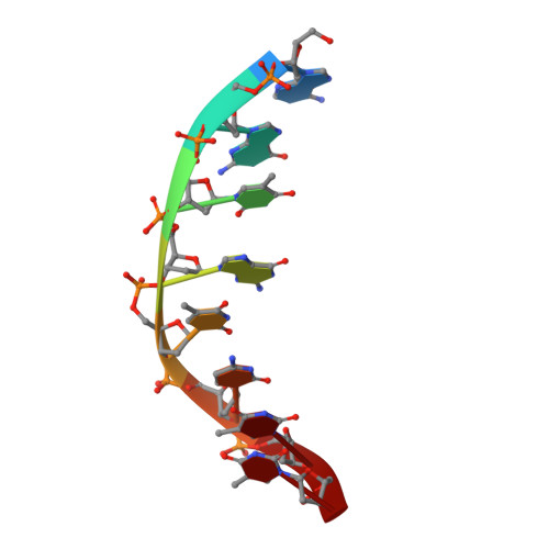
Crystal structure of the Rous sarcoma virus intasome.

Yin, Z.Shi, K.Banerjee, S.Pandey, K.K.Bera, S.Grandgenett, D.P.Aihara, H.

(2016) Nature 530: 362-366

  • DOI: https://doi.org/10.1038/nature16950
  • Primary Citation of Related Structures:  
    5EJK

  • PubMed Abstract: 

    Integration of the reverse-transcribed viral DNA into the host genome is an essential step in the life cycle of retroviruses. Retrovirus integrase catalyses insertions of both ends of the linear viral DNA into a host chromosome. Integrase from HIV-1 and closely related retroviruses share the three-domain organization, consisting of a catalytic core domain flanked by amino- and carboxy-terminal domains essential for the concerted integration reaction. Although structures of the tetrameric integrase-DNA complexes have been reported for integrase from prototype foamy virus featuring an additional DNA-binding domain and longer interdomain linkers, the architecture of a canonical three-domain integrase bound to DNA remained elusive. Here we report a crystal structure of the three-domain integrase from Rous sarcoma virus in complex with viral and target DNAs. The structure shows an octameric assembly of integrase, in which a pair of integrase dimers engage viral DNA ends for catalysis while another pair of non-catalytic integrase dimers bridge between the two viral DNA molecules and help capture target DNA. The individual domains of the eight integrase molecules play varying roles to hold the complex together, making an extensive network of protein-DNA and protein-protein contacts that show both conserved and distinct features compared with those observed for prototype foamy virus integrase. Our work highlights the diversity of retrovirus intasome assembly and provides insights into the mechanisms of integration by HIV-1 and related retroviruses.


  • Organizational Affiliation

    Department of Biochemistry, Molecular Biology and Biophysics, University of Minnesota, Minneapolis, Minnesota 55455, USA.


Macromolecules

Find similar proteins by:  (by identity cutoff)  |  3D Structure
Entity ID: 1
MoleculeChains Sequence LengthOrganismDetailsImage
Gag-Pro-Pol polyprotein
A, B, C, D, E
A, B, C, D, E, F, G, H
270Rous sarcoma virus - Prague CMutation(s): 7 
Gene Names: gag-pro-pol
EC: 3.4.23 (PDB Primary Data), 2.7.7.49 (PDB Primary Data), 2.7.7.7 (PDB Primary Data), 3.1.26.4 (PDB Primary Data), 2.7.7 (PDB Primary Data), 3.1 (PDB Primary Data)
UniProt
Find proteins for P03354 (Rous sarcoma virus subgroup C (strain Prague))
Explore P03354 
Go to UniProtKB:  P03354
Entity Groups  
Sequence Clusters30% Identity50% Identity70% Identity90% Identity95% Identity100% Identity
UniProt GroupP03354
Sequence Annotations
Expand
  • Reference Sequence
Find similar nucleic acids by:  (by identity cutoff)  |  3D Structure
Entity ID: 2
MoleculeChains LengthOrganismImage
RSV Integrase
I, L
42Rous sarcoma virus (strain Schmidt-Ruppin E)
Sequence Annotations
Expand
  • Reference Sequence

Find similar nucleic acids by:  Sequence   |   3D Structure  

Entity ID: 3
MoleculeChains LengthOrganismImage
DNA (5'-D(*AP*AP*TP*GP*TP*TP*GP*TP*CP*TP*TP*AP*TP*GP*CP*AP*AP*TP*AP*CP*TP*C)-3')
J, M
22Rous sarcoma virus (strain Schmidt-Ruppin E)
Sequence Annotations
Expand
  • Reference Sequence

Find similar nucleic acids by:  Sequence   |   3D Structure  

Entity ID: 4
MoleculeChains LengthOrganismImage
DNA (5'-D(*CP*TP*TP*CP*TP*CP*TP*C)-3')
K, N
8Homo sapiens
Sequence Annotations
Expand
  • Reference Sequence

Find similar nucleic acids by:  Sequence   |   3D Structure  

Entity ID: 5
MoleculeChains LengthOrganismImage
DNA (5'-D(*AP*GP*TP*GP*TP*CP*TP*T)-3')O [auth k],
P [auth n]
8Homo sapiens
Sequence Annotations
Expand
  • Reference Sequence
Small Molecules
Ligands 2 Unique
IDChains Name / Formula / InChI Key2D Diagram3D Interactions
W
Query on W

Download Ideal Coordinates CCD File 
AA [auth B]
AB [auth F]
BA [auth B]
BB [auth F]
CA [auth B]
AA [auth B],
AB [auth F],
BA [auth B],
BB [auth F],
CA [auth B],
CB [auth F],
DA [auth B],
DB [auth F],
EA [auth B],
EB [auth F],
FA [auth B],
FB [auth G],
GA [auth B],
HA [auth B],
IA [auth B],
JA [auth B],
KA [auth B],
LA [auth B],
MA [auth B],
NA [auth B],
OA [auth B],
PA [auth B],
R [auth A],
S [auth A],
T [auth A],
U [auth A],
UA [auth F],
VA [auth F],
W [auth B],
WA [auth F],
X [auth B],
XA [auth F],
Y [auth B],
YA [auth F],
Z [auth B],
ZA [auth F]
TUNGSTEN ION
W
FZFRVZDLZISPFJ-UHFFFAOYSA-N
ZN
Query on ZN

Download Ideal Coordinates CCD File 
GB [auth G]
HB [auth H]
Q [auth A]
QA [auth C]
RA [auth D]
GB [auth G],
HB [auth H],
Q [auth A],
QA [auth C],
RA [auth D],
SA [auth E],
TA [auth F],
V [auth B]
ZINC ION
Zn
PTFCDOFLOPIGGS-UHFFFAOYSA-N
Modified Residues  1 Unique
IDChains TypeFormula2D DiagramParent
MSE
Query on MSE
A, B, C, D, E
A, B, C, D, E, F, G, H
L-PEPTIDE LINKINGC5 H11 N O2 SeMET
Experimental Data & Validation

Experimental Data

  • Method: X-RAY DIFFRACTION
  • Resolution: 3.80 Å
  • R-Value Free: 0.294 
  • R-Value Work: 0.254 
  • R-Value Observed: 0.256 
  • Space Group: P 1 21 1
Unit Cell:
Length ( Å )Angle ( ˚ )
a = 124.936α = 90
b = 157.854β = 110.94
c = 126.583γ = 90
Software Package:
Software NamePurpose
PHENIXrefinement
XDSdata reduction
XSCALEdata scaling
PHASERphasing

Structure Validation

View Full Validation Report


Entry History 

Deposition Data

Revision History  (Full details and data files)

  • Version 1.0: 2016-02-17
    Type: Initial release
  • Version 1.1: 2023-09-27
    Changes: Data collection, Database references, Derived calculations, Refinement description
  • Version 1.2: 2023-11-15
    Changes: Data collection