5E9S

Crystal structure of substrate-bound glutamate transporter homologue GltTk


Experimental Data Snapshot

  • Method: X-RAY DIFFRACTION
  • Resolution: 2.80 Å
  • R-Value Free: 0.242 
  • R-Value Work: 0.213 
  • R-Value Observed: 0.214 

wwPDB Validation   3D Report Full Report


Ligand Structure Quality Assessment 


This is version 1.3 of the entry. See complete history

Re-refinement Note

A newer entry is available that reflects an alternative modeling of the original data: 8AFA


Literature

Coupled binding mechanism of three sodium ions and aspartate in the glutamate transporter homologue GltTk.

Guskov, A.Jensen, S.Faustino, I.Marrink, S.J.Slotboom, D.J.

(2016) Nat Commun 7: 13420-13420

  • DOI: https://doi.org/10.1038/ncomms13420
  • Primary Citation of Related Structures:  
    5E9S

  • PubMed Abstract: 

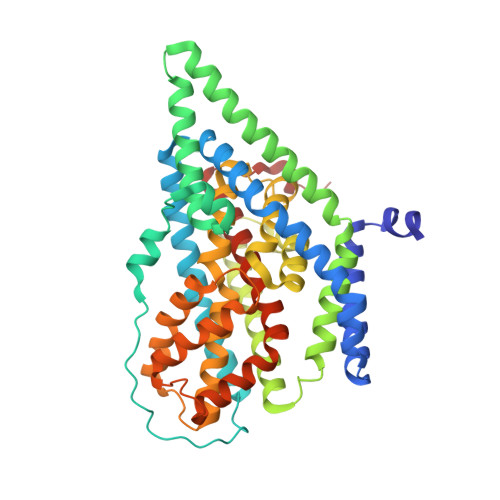
    Glutamate transporters catalyse the thermodynamically unfavourable transport of anionic amino acids across the cell membrane by coupling it to the downhill transport of cations. This coupling mechanism is still poorly understood, in part because the available crystal structures of these transporters are of relatively low resolution. Here we solve crystal structures of the archaeal transporter Glt Tk in the presence and absence of aspartate and use molecular dynamics simulations and binding assays to show how strict coupling between the binding of three sodium ions and aspartate takes place.


  • Organizational Affiliation

    University of Groningen, Groningen Biomolecular Sciences and Biotechnology Institute, Nijenborgh 4, 9747 AG Groningen, The Netherlands.


Macromolecules
Find similar proteins by:  (by identity cutoff)  |  3D Structure
Entity ID: 1
MoleculeChains Sequence LengthOrganismDetailsImage
Proton/glutamate symporter, SDF family
A, B, C
438Thermococcus kodakarensisMutation(s): 0 
Gene Names: TK0986
Membrane Entity: Yes 
UniProt
Find proteins for Q5JID0 (Thermococcus kodakarensis (strain ATCC BAA-918 / JCM 12380 / KOD1))
Explore Q5JID0 
Go to UniProtKB:  Q5JID0
Entity Groups  
Sequence Clusters30% Identity50% Identity70% Identity90% Identity95% Identity100% Identity
UniProt GroupQ5JID0
Sequence Annotations
Expand
  • Reference Sequence
Small Molecules
Ligands 7 Unique
IDChains Name / Formula / InChI Key2D Diagram3D Interactions
DMU
Query on DMU

Download Ideal Coordinates CCD File 
CB [auth C],
DA [auth B],
H [auth A]
DECYL-BETA-D-MALTOPYRANOSIDE
C22 H42 O11
WOQQAWHSKSSAGF-WXFJLFHKSA-N
P6G
Query on P6G

Download Ideal Coordinates CCD File 
QB [auth C],
RB [auth C],
WA [auth B],
XA [auth B],
Y [auth A]
HEXAETHYLENE GLYCOL
C12 H26 O7
IIRDTKBZINWQAW-UHFFFAOYSA-N
1PE
Query on 1PE

Download Ideal Coordinates CCD File 
DB [auth C]
EA [auth B]
EB [auth C]
FA [auth B]
GA [auth B]
DB [auth C],
EA [auth B],
EB [auth C],
FA [auth B],
GA [auth B],
I [auth A],
J [auth A]
PENTAETHYLENE GLYCOL
C10 H22 O6
JLFNLZLINWHATN-UHFFFAOYSA-N
PGE
Query on PGE

Download Ideal Coordinates CCD File 
OB [auth C]
PB [auth C]
U [auth A]
V [auth A]
VA [auth B]
OB [auth C],
PB [auth C],
U [auth A],
V [auth A],
VA [auth B],
W [auth A],
X [auth A]
TRIETHYLENE GLYCOL
C6 H14 O4
ZIBGPFATKBEMQZ-UHFFFAOYSA-N
ASP
Query on ASP

Download Ideal Coordinates CCD File 
D [auth A],
YA [auth C],
Z [auth B]
ASPARTIC ACID
C4 H7 N O4
CKLJMWTZIZZHCS-REOHCLBHSA-N
PEG
Query on PEG

Download Ideal Coordinates CCD File 
FB [auth C]
GB [auth C]
HA [auth B]
HB [auth C]
IA [auth B]
FB [auth C],
GB [auth C],
HA [auth B],
HB [auth C],
IA [auth B],
IB [auth C],
JA [auth B],
JB [auth C],
K [auth A],
KA [auth B],
KB [auth C],
L [auth A],
LA [auth B],
LB [auth C],
M [auth A],
MA [auth B],
MB [auth C],
N [auth A],
NA [auth B],
NB [auth C],
O [auth A],
OA [auth B],
P [auth A],
PA [auth B],
Q [auth A],
QA [auth B],
R [auth A],
RA [auth B],
S [auth A],
SA [auth B],
T [auth A],
TA [auth B],
UA [auth B]
DI(HYDROXYETHYL)ETHER
C4 H10 O3
MTHSVFCYNBDYFN-UHFFFAOYSA-N
NA
Query on NA

Download Ideal Coordinates CCD File 
AA [auth B]
AB [auth C]
BA [auth B]
BB [auth C]
CA [auth B]
AA [auth B],
AB [auth C],
BA [auth B],
BB [auth C],
CA [auth B],
E [auth A],
F [auth A],
G [auth A],
ZA [auth C]
SODIUM ION
Na
FKNQFGJONOIPTF-UHFFFAOYSA-N
Experimental Data & Validation

Experimental Data

  • Method: X-RAY DIFFRACTION
  • Resolution: 2.80 Å
  • R-Value Free: 0.242 
  • R-Value Work: 0.213 
  • R-Value Observed: 0.214 
  • Space Group: P 32 2 1
Unit Cell:
Length ( Å )Angle ( ˚ )
a = 117.52α = 90
b = 117.52β = 90
c = 310.59γ = 120
Software Package:
Software NamePurpose
PHENIXrefinement
XDSdata reduction
XSCALEdata scaling
PHASERphasing

Structure Validation

View Full Validation Report


Ligand Structure Quality Assessment 


Entry History & Funding Information

Deposition Data


Funding OrganizationLocationGrant Number
NWONetherlands865.11.001

Revision History  (Full details and data files)

  • Version 1.0: 2016-09-28
    Type: Initial release
  • Version 1.1: 2016-11-16
    Changes: Database references
  • Version 1.2: 2016-11-23
    Changes: Database references
  • Version 1.3: 2024-01-10
    Changes: Data collection, Database references, Refinement description