5E5C

Crystal structure of dihydropyrimidinase from Pseudomonas aeruginosa PAO1


Experimental Data Snapshot

  • Method: X-RAY DIFFRACTION
  • Resolution: 2.10 Å
  • R-Value Free: 0.215 
  • R-Value Work: 0.170 
  • R-Value Observed: 0.172 

wwPDB Validation   3D Report Full Report


This is version 1.3 of the entry. See complete history


Literature

Crystal structure of dihydropyrimidinase from Pseudomonas aeruginosa PAO1: Insights into the molecular basis of formation of a dimer

Tzeng, C.T.Huang, Y.H.Huang, C.Y.

(2016) Biochem Biophys Res Commun 478: 1449-1455

  • DOI: https://doi.org/10.1016/j.bbrc.2016.08.144
  • Primary Citation of Related Structures:  
    5E5C

  • PubMed Abstract: 

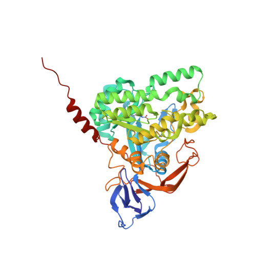
    Dihydropyrimidinase, a tetrameric metalloenzyme, is a member of the cyclic amidohydrolase family, which also includes allantoinase, dihydroorotase, hydantoinase, and imidase. In this paper, we report the crystal structure of dihydropyrimidinase from Pseudomonas aeruginosa PAO1 at 2.1 Å resolution. The structure of P. aeruginosa dihydropyrimidinase reveals a classic (β/α)8-barrel structure core embedding the catalytic dimetal center and a β-sandwich domain, which is commonly found in the architecture of dihydropyrimidinases. In contrast to all dihydropyrimidinases, P. aeruginosa dihydropyrimidinase forms a dimer, rather than a tetramer, both in the crystalline state and in the solution. Basing on sequence analysis and structural comparison of the C-terminal region and the dimer-dimer interface between P. aeruginosa dihydropyrimidinase and Thermus sp. dihydropyrimidinase, we propose a working model to explain why this enzyme cannot be a tetramer.


  • Organizational Affiliation

    School of Biomedical Sciences, Chung Shan Medical University, No. 110, Sec. 1, Chien-Kuo N. Rd., Taichung City, Taiwan.


Macromolecules
Find similar proteins by:  (by identity cutoff)  |  3D Structure
Entity ID: 1
MoleculeChains Sequence LengthOrganismDetailsImage
D-hydantoinase/dihydropyrimidinaseA,
B [auth C]
485Pseudomonas aeruginosa PAO1Mutation(s): 0 
Gene Names: dhtPA0441
EC: 3.5.2.2
UniProt
Find proteins for Q9I676 (Pseudomonas aeruginosa (strain ATCC 15692 / DSM 22644 / CIP 104116 / JCM 14847 / LMG 12228 / 1C / PRS 101 / PAO1))
Explore Q9I676 
Go to UniProtKB:  Q9I676
Entity Groups  
Sequence Clusters30% Identity50% Identity70% Identity90% Identity95% Identity100% Identity
UniProt GroupQ9I676
Sequence Annotations
Expand
  • Reference Sequence
Experimental Data & Validation

Experimental Data

  • Method: X-RAY DIFFRACTION
  • Resolution: 2.10 Å
  • R-Value Free: 0.215 
  • R-Value Work: 0.170 
  • R-Value Observed: 0.172 
  • Space Group: P 31 2 1
Unit Cell:
Length ( Å )Angle ( ˚ )
a = 112.134α = 90
b = 112.134β = 90
c = 161.05γ = 120
Software Package:
Software NamePurpose
REFMACrefinement
HKL-2000data scaling
HKL-2000data reduction
MOLREPphasing

Structure Validation

View Full Validation Report


Entry History 

Deposition Data

Revision History  (Full details and data files)

  • Version 1.0: 2016-09-21
    Type: Initial release
  • Version 1.1: 2016-09-28
    Changes: Database references
  • Version 1.2: 2023-11-08
    Changes: Data collection, Database references, Derived calculations, Refinement description
  • Version 1.3: 2023-11-15
    Changes: Data collection