5E30

Crystal structure of H5 hemagglutinin Q226L mutant from the influenza virus A/duck/Egypt/10185SS/2010 (H5N1) with LSTc


Experimental Data Snapshot

  • Method: X-RAY DIFFRACTION
  • Resolution: 2.70 Å
  • R-Value Free: 0.249 
  • R-Value Work: 0.202 
  • R-Value Observed: 0.204 

wwPDB Validation   3D Report Full Report


Ligand Structure Quality Assessment 


This is version 2.1 of the entry. See complete history


Literature

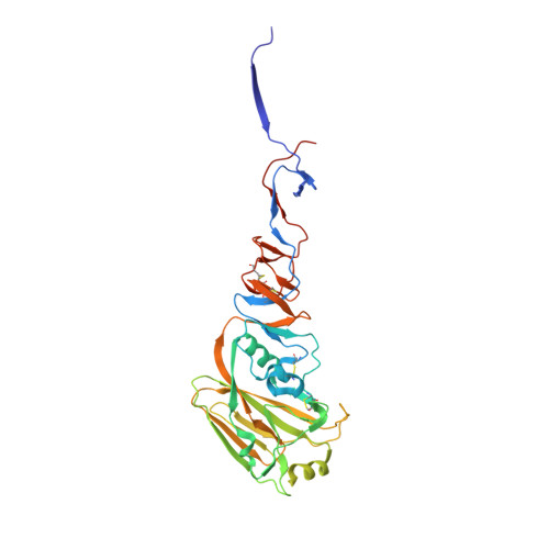
Structural Basis for a Switch in Receptor Binding Specificity of Two H5N1 Hemagglutinin Mutants.

Zhu, X.Viswanathan, K.Raman, R.Yu, W.Sasisekharan, R.Wilson, I.A.

(2015) Cell Rep 13: 1683-1691

  • DOI: https://doi.org/10.1016/j.celrep.2015.10.027
  • Primary Citation of Related Structures:  
    5E2Y, 5E2Z, 5E30, 5E32, 5E34, 5E35

  • PubMed Abstract: 

    Avian H5N1 influenza viruses continue to spread in wild birds and domestic poultry with sporadic infection in humans. Receptor binding specificity changes are a prerequisite for H5N1 viruses and other zoonotic viruses to be transmitted among humans. Previous reported hemagglutinin (HA) mutants from ferret-transmissible H5N1 viruses of A/Vietnam/1203/2004 and A/Indonesia/5/2005 showed slightly increased, but still very weak, binding to human receptors. From mutagenesis and glycan array studies, we previously identified two H5N1 HA mutants that could more effectively switch receptor specificity to human-like α2-6-linked sialosides with avidity comparable to wild-type H5 HA binding to avian-like α2-3-linked sialosides. Here, crystal structures of these two H5 HA mutants free and in complex with human and avian glycan receptor analogs reveal the structural basis for their preferential binding to human receptors. These findings suggest continuous surveillance should be maintained to monitor and assess human-to-human transmission potential of H5N1 viruses.


  • Organizational Affiliation

    Department of Integrative Structural and Computational Biology, The Scripps Research Institute, La Jolla, CA 92037, USA.


Macromolecules
Find similar proteins by:  (by identity cutoff)  |  3D Structure
Entity ID: 1
MoleculeChains Sequence LengthOrganismDetailsImage
HemagglutininA [auth C],
C [auth A],
E
333Influenza A virus (A/duck/Egypt/10185SS/2010(H5N1))Mutation(s): 1 
Gene Names: HA
UniProt
Find proteins for G8IPF0 (Influenza A virus)
Explore G8IPF0 
Go to UniProtKB:  G8IPF0
Entity Groups  
Sequence Clusters30% Identity50% Identity70% Identity90% Identity95% Identity100% Identity
UniProt GroupG8IPF0
Sequence Annotations
Expand
  • Reference Sequence
Find similar proteins by:  (by identity cutoff)  |  3D Structure
Entity ID: 2
MoleculeChains Sequence LengthOrganismDetailsImage
HemagglutininB [auth D],
D [auth B],
F
180Influenza A virus (A/duck/Egypt/10185SS/2010(H5N1))Mutation(s): 0 
Gene Names: HA
UniProt
Find proteins for G8IPF0 (Influenza A virus)
Explore G8IPF0 
Go to UniProtKB:  G8IPF0
Entity Groups  
Sequence Clusters30% Identity50% Identity70% Identity90% Identity95% Identity100% Identity
UniProt GroupG8IPF0
Sequence Annotations
Expand
  • Reference Sequence
Oligosaccharides

Help

Entity ID: 3
MoleculeChains Length2D Diagram Glycosylation3D Interactions
beta-L-fucopyranose-(1-3)-[2-acetamido-2-deoxy-beta-D-glucopyranose-(1-4)][alpha-L-fucopyranose-(1-6)]2-acetamido-2-deoxy-beta-D-glucopyranose
G
4N-Glycosylation
Glycosylation Resources
GlyTouCan:  G62810GW
GlyCosmos:  G62810GW
Entity ID: 4
MoleculeChains Length2D Diagram Glycosylation3D Interactions
N-acetyl-alpha-neuraminic acid-(2-6)-beta-D-galactopyranose-(1-4)-2-acetamido-2-deoxy-beta-D-glucopyranose-(1-3)-beta-D-galactopyranose
H, J, L
4N/A
Glycosylation Resources
GlyTouCan:  G03509EL
GlyCosmos:  G03509EL
GlyGen:  G03509EL
Entity ID: 5
MoleculeChains Length2D Diagram Glycosylation3D Interactions
beta-L-fucopyranose-(1-3)-[2-acetamido-2-deoxy-beta-D-glucopyranose-(1-4)]2-acetamido-2-deoxy-beta-D-glucopyranose
I, K
3N-Glycosylation
Glycosylation Resources
GlyTouCan:  G46874CY
GlyCosmos:  G46874CY
GlyGen:  G46874CY
Small Molecules
Ligands 1 Unique
IDChains Name / Formula / InChI Key2D Diagram3D Interactions
NAG
Query on NAG

Download Ideal Coordinates CCD File 
M [auth C],
N [auth E]
2-acetamido-2-deoxy-beta-D-glucopyranose
C8 H15 N O6
OVRNDRQMDRJTHS-FMDGEEDCSA-N
Experimental Data & Validation

Experimental Data

  • Method: X-RAY DIFFRACTION
  • Resolution: 2.70 Å
  • R-Value Free: 0.249 
  • R-Value Work: 0.202 
  • R-Value Observed: 0.204 
  • Space Group: P 1 21 1
Unit Cell:
Length ( Å )Angle ( ˚ )
a = 69.901α = 90
b = 228.524β = 114.29
c = 70.682γ = 90
Software Package:
Software NamePurpose
PHENIXrefinement
HKL-2000data reduction
HKL-2000data scaling
PHASERphasing

Structure Validation

View Full Validation Report


Ligand Structure Quality Assessment 


Entry History 

Deposition Data

Revision History  (Full details and data files)

  • Version 1.0: 2015-12-02
    Type: Initial release
  • Version 1.1: 2015-12-09
    Changes: Database references
  • Version 2.0: 2020-07-29
    Type: Remediation
    Reason: Carbohydrate remediation
    Changes: Atomic model, Data collection, Derived calculations, Structure summary
  • Version 2.1: 2023-09-27
    Changes: Data collection, Database references, Derived calculations, Refinement description, Structure summary