5DP8

Crystal Structure of EV71 3C Proteinase in complex with compound 8


Experimental Data Snapshot

  • Method: X-RAY DIFFRACTION
  • Resolution: 2.40 Å
  • R-Value Free: 0.276 
  • R-Value Work: 0.208 
  • R-Value Observed: 0.211 

wwPDB Validation   3D Report Full Report


Ligand Structure Quality Assessment 


This is version 1.3 of the entry. See complete history


Literature

Fragment-wise design of inhibitors to 3C proteinase from enterovirus 71

Wu, C.Zhang, L.Li, P.Cai, Q.Peng, X.Yin, K.Chen, X.Ren, H.Zhong, S.Weng, Y.Guan, Y.Chen, S.Wu, J.Li, J.Lin, T.

(2016) Biochim Biophys Acta 1860: 1299-1307

  • DOI: https://doi.org/10.1016/j.bbagen.2016.03.017
  • Primary Citation of Related Structures:  
    5DP3, 5DP4, 5DP5, 5DP6, 5DP7, 5DP8, 5DP9, 5DPA

  • PubMed Abstract: 

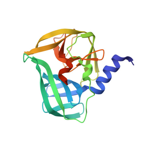
    Enterovirus 71 (EV71) is a causative agent of hand, foot and mouth disease (HFMD), which can spread its infection to central nervous and other systems with severe consequence. A key factor in the replication of EV71 is its 3C proteinase (3C(pro)), a significant drug target. Peptidomimetics were employed as inhibitors of this enzyme for developing antivirals. However, the peptide bonds in these peptidomimetics are a source of low bioavailability due to their susceptibility to protease digestion. To produce non-peptidomimetic inhibitors by replacing these peptide bonds, it would be important to gain better understanding on the contribution of each component to the interaction and potency.


  • Organizational Affiliation

    The First Affiliated Hospital of Xiamen University, Xiamen, Fujian, China; State Key Laboratory of Cellular Stress Biology, Innovation Center for Cell Signaling Network, State-Province Joint Engineering Laboratory of Targeted Drugs from Natural Products, School of Life Sciences, Xiamen University, Xiamen, Fujian, China.


Macromolecules
Find similar proteins by:  (by identity cutoff)  |  3D Structure
Entity ID: 1
MoleculeChains Sequence LengthOrganismDetailsImage
3C proteinase192Enterovirus A71Mutation(s): 0 
UniProt
Find proteins for A9XG43 (Human enterovirus 71)
Explore A9XG43 
Go to UniProtKB:  A9XG43
Entity Groups  
Sequence Clusters30% Identity50% Identity70% Identity90% Identity95% Identity100% Identity
UniProt GroupA9XG43
Sequence Annotations
Expand
  • Reference Sequence
Small Molecules
Ligands 1 Unique
IDChains Name / Formula / InChI Key2D Diagram3D Interactions
5ED
Query on 5ED

Download Ideal Coordinates CCD File 
B [auth A]ethyl (2Z,4S)-4-[(N-{[(2-cyclopropylethyl)amino](oxo)acetyl}-L-phenylalanyl)amino]-5-[(3S)-2-oxopyrrolidin-3-yl]pent-2-enoate
C27 H36 N4 O6
HYAHTJFZAHAFHT-BDTNDASRSA-N
Experimental Data & Validation

Experimental Data

  • Method: X-RAY DIFFRACTION
  • Resolution: 2.40 Å
  • R-Value Free: 0.276 
  • R-Value Work: 0.208 
  • R-Value Observed: 0.211 
  • Space Group: C 2 2 21
Unit Cell:
Length ( Å )Angle ( ˚ )
a = 63.945α = 90
b = 64.976β = 90
c = 75.606γ = 90
Software Package:
Software NamePurpose
REFMACrefinement
HKL-2000data scaling
PDB_EXTRACTdata extraction

Structure Validation

View Full Validation Report


Ligand Structure Quality Assessment 


Entry History 

Deposition Data

Revision History  (Full details and data files)

  • Version 1.0: 2016-03-30
    Type: Initial release
  • Version 1.1: 2016-04-06
    Changes: Database references
  • Version 1.2: 2017-09-27
    Changes: Data collection, Database references, Derived calculations
  • Version 1.3: 2023-11-08
    Changes: Data collection, Database references, Refinement description