5D6P

Crystal structure of the ATP binding domain of S. aureus GyrB complexed with a ligand


Experimental Data Snapshot

  • Method: X-RAY DIFFRACTION
  • Resolution: 2.05 Å
  • R-Value Free: 0.257 
  • R-Value Work: 0.208 
  • R-Value Observed: 0.211 

wwPDB Validation   3D Report Full Report


Ligand Structure Quality Assessment 


This is version 1.1 of the entry. See complete history


Literature

Discovery of Azaindole Ureas as a Novel Class of Bacterial Gyrase B Inhibitors.

Zhang, J.Yang, Q.Cross, J.B.Romero, J.A.Poutsiaka, K.M.Epie, F.Bevan, D.Wang, B.Zhang, Y.Chavan, A.Zhang, X.Moy, T.Daniel, A.Nguyen, K.Chamberlain, B.Carter, N.Shotwell, J.Silverman, J.Metcalf, C.A.Ryan, D.Lippa, B.Dolle, R.E.

(2015) J Med Chem 58: 8503-8512

  • DOI: https://doi.org/10.1021/acs.jmedchem.5b00961
  • Primary Citation of Related Structures:  
    5D6P, 5D6Q, 5D7C

  • PubMed Abstract: 

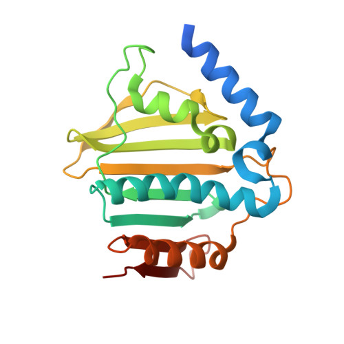
    The emergence and spread of multidrug resistant bacteria are widely believed to endanger human health. New drug targets and lead compounds exempt from cross-resistance with existing drugs are urgently needed. We report on the discovery of azaindole ureas as a novel class of bacterial gyrase B inhibitors and detail the story of their evolution from a de novo design hit based on structure-based drug design. These inhibitors show potent minimum inhibitory concentrations against fluoroquinolone resistant MRSA and other Gram-positive bacteria.


  • Organizational Affiliation

    Cubist Pharmaceuticals Inc. , Lexington, Massachusetts 02421, United States.


Macromolecules
Find similar proteins by:  (by identity cutoff)  |  3D Structure
Entity ID: 1
MoleculeChains Sequence LengthOrganismDetailsImage
DNA gyrase subunit B
A, B
212Staphylococcus aureusMutation(s): 0 
Gene Names: gyrB
EC: 5.99.1.3
UniProt
Find proteins for P0A0K8 (Staphylococcus aureus)
Explore P0A0K8 
Go to UniProtKB:  P0A0K8
Entity Groups  
Sequence Clusters30% Identity50% Identity70% Identity90% Identity95% Identity100% Identity
UniProt GroupP0A0K8
Sequence Annotations
Expand
  • Reference Sequence
Small Molecules
Binding Affinity Annotations 
IDSourceBinding Affinity
57U Binding MOAD:  5D6P IC50: 2500 (nM) from 1 assay(s)
BindingDB:  5D6P IC50: 2500 (nM) from 1 assay(s)
Experimental Data & Validation

Experimental Data

  • Method: X-RAY DIFFRACTION
  • Resolution: 2.05 Å
  • R-Value Free: 0.257 
  • R-Value Work: 0.208 
  • R-Value Observed: 0.211 
  • Space Group: C 1 2 1
Unit Cell:
Length ( Å )Angle ( ˚ )
a = 144.539α = 90
b = 55.556β = 99.73
c = 51.113γ = 90
Software Package:
Software NamePurpose
REFMACrefinement
d*TREKdata reduction
d*TREKdata scaling
MOLREPphasing
PDB_EXTRACTdata extraction

Structure Validation

View Full Validation Report


Ligand Structure Quality Assessment 


Entry History 

Revision History  (Full details and data files)

  • Version 1.0: 2015-11-25
    Type: Initial release
  • Version 1.1: 2023-09-27
    Changes: Data collection, Database references, Derived calculations, Refinement description