5CL5

Alkylpurine DNA glycosylase AlkD bound to DNA containing a 3-methyladenine analog or DNA containing an abasic site and a free nucleobase (51% substrate/49% product at 48 hours)


Experimental Data Snapshot

  • Method: X-RAY DIFFRACTION
  • Resolution: 1.57 Å
  • R-Value Free: 0.189 
  • R-Value Work: 0.158 
  • R-Value Observed: 0.159 

wwPDB Validation   3D Report Full Report


This is version 1.5 of the entry. See complete history


Literature

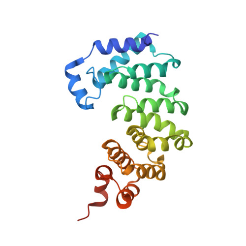
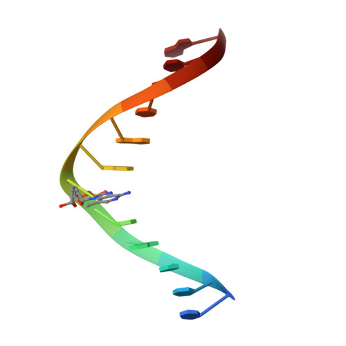
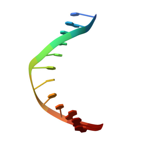
The DNA glycosylase AlkD uses a non-base-flipping mechanism to excise bulky lesions.

Mullins, E.A.Shi, R.Parsons, Z.D.Yuen, P.K.David, S.S.Igarashi, Y.Eichman, B.F.

(2015) Nature 527: 254-258

  • DOI: https://doi.org/10.1038/nature15728
  • Primary Citation of Related Structures:  
    5CL3, 5CL4, 5CL5, 5CL6, 5CL7, 5CL8, 5CL9, 5CLA, 5CLB, 5CLC, 5CLD, 5CLE

  • PubMed Abstract: 

    Threats to genomic integrity arising from DNA damage are mitigated by DNA glycosylases, which initiate the base excision repair pathway by locating and excising aberrant nucleobases. How these enzymes find small modifications within the genome is a current area of intensive research. A hallmark of these and other DNA repair enzymes is their use of base flipping to sequester modified nucleotides from the DNA helix and into an active site pocket. Consequently, base flipping is generally regarded as an essential aspect of lesion recognition and a necessary precursor to base excision. Here we present the first, to our knowledge, DNA glycosylase mechanism that does not require base flipping for either binding or catalysis. Using the DNA glycosylase AlkD from Bacillus cereus, we crystallographically monitored excision of an alkylpurine substrate as a function of time, and reconstructed the steps along the reaction coordinate through structures representing substrate, intermediate and product complexes. Instead of directly interacting with the damaged nucleobase, AlkD recognizes aberrant base pairs through interactions with the phosphoribose backbone, while the lesion remains stacked in the DNA duplex. Quantum mechanical calculations revealed that these contacts include catalytic charge-dipole and CH-π interactions that preferentially stabilize the transition state. We show in vitro and in vivo how this unique means of recognition and catalysis enables AlkD to repair large adducts formed by yatakemycin, a member of the duocarmycin family of antimicrobial natural products exploited in bacterial warfare and chemotherapeutic trials. Bulky adducts of this or any type are not excised by DNA glycosylases that use a traditional base-flipping mechanism. Hence, these findings represent a new model for DNA repair and provide insights into catalysis of base excision.


  • Organizational Affiliation

    Department of Biological Sciences and Center for Structural Biology, Vanderbilt University, Nashville, Tennessee 37232, USA.


Macromolecules

Find similar proteins by:  (by identity cutoff)  |  3D Structure
Entity ID: 1
MoleculeChains Sequence LengthOrganismDetailsImage
AlkD241Bacillus cereusMutation(s): 0 
Gene Names: IKE_03968
EC: 3.2.2
UniProt
Find proteins for Q816E8 (Bacillus cereus (strain ATCC 14579 / DSM 31 / CCUG 7414 / JCM 2152 / NBRC 15305 / NCIMB 9373 / NCTC 2599 / NRRL B-3711))
Explore Q816E8 
Go to UniProtKB:  Q816E8
Entity Groups  
Sequence Clusters30% Identity50% Identity70% Identity90% Identity95% Identity100% Identity
UniProt GroupQ816E8
Sequence Annotations
Expand
  • Reference Sequence

Find similar nucleic acids by:  Sequence   |   3D Structure  

Entity ID: 2
MoleculeChains LengthOrganismImage
DNA (5'-D(*CP*CP*CP*GP*AP*(DZM)P*AP*GP*TP*CP*CP*G)-3')12synthetic construct
Sequence Annotations
Expand
  • Reference Sequence

Find similar nucleic acids by:  Sequence   |   3D Structure  

Entity ID: 3
MoleculeChains LengthOrganismImage
DNA (5'-D(*CP*GP*GP*AP*CP*TP*TP*TP*CP*GP*GP*G)-3')12synthetic construct
Sequence Annotations
Expand
  • Reference Sequence
Small Molecules
Ligands 1 Unique
IDChains Name / Formula / InChI Key2D Diagram3D Interactions
54K
Query on 54K

Download Ideal Coordinates CCD File 
D [auth B]7-methyl-3H-imidazo[4,5-c]pyridin-4-amine
C7 H8 N4
VUSDRZRNZRBERR-UHFFFAOYSA-N
Experimental Data & Validation

Experimental Data

  • Method: X-RAY DIFFRACTION
  • Resolution: 1.57 Å
  • R-Value Free: 0.189 
  • R-Value Work: 0.158 
  • R-Value Observed: 0.159 
  • Space Group: P 1 21 1
Unit Cell:
Length ( Å )Angle ( ˚ )
a = 38.341α = 90
b = 93.4β = 112.85
c = 48.117γ = 90
Software Package:
Software NamePurpose
PHENIXrefinement
HKL-2000data scaling
PDB_EXTRACTdata extraction
HKL-2000data reduction
PHENIXphasing
Cootmodel building

Structure Validation

View Full Validation Report


Entry History & Funding Information

Deposition Data


Funding OrganizationLocationGrant Number
National Science Foundation (NSF, United States)United StatesMCB-1122098

Revision History  (Full details and data files)

  • Version 1.0: 2015-10-28
    Type: Initial release
  • Version 1.1: 2015-11-18
    Changes: Database references
  • Version 1.2: 2015-11-25
    Changes: Database references
  • Version 1.3: 2017-09-13
    Changes: Author supporting evidence, Database references, Derived calculations
  • Version 1.4: 2019-11-27
    Changes: Author supporting evidence
  • Version 1.5: 2023-09-27
    Changes: Data collection, Database references, Refinement description