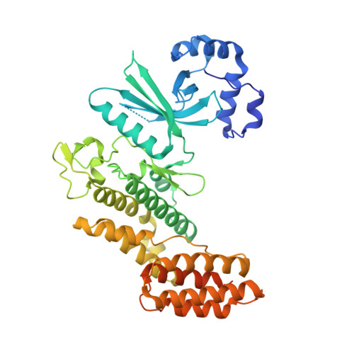
5CKW

Crystal structure of LegK4_AMPPNP Kinase


Experimental Data Snapshot

  • Method: X-RAY DIFFRACTION
  • Resolution: 2.49 Å
  • R-Value Free: 0.258 
  • R-Value Work: 0.204 
  • R-Value Observed: 0.207 

wwPDB Validation   3D Report Full Report


Ligand Structure Quality Assessment 


This is version 1.1 of the entry. See complete history


Literature

The structure of Legionella pneumophila LegK4 type four secretion system (T4SS) effector reveals a novel dimeric eukaryotic-like kinase.

Flayhan, A.Berge, C.Bailo, N.Doublet, P.Bayliss, R.Terradot, L.

(2015) Sci Rep 5: 14602-14602

  • DOI: https://doi.org/10.1038/srep14602
  • Primary Citation of Related Structures:  
    5CKW, 5CLR

  • PubMed Abstract: 

    Bacterial pathogens subvert signalling pathways to promote invasion and/or replication into the host. LegK1-4 proteins are eukaryotic-like serine/threonine kinases that are translocated by the Dot/Icm type IV secretion system (T4SS) of several Legionella pneumophila strains. We present the crystal structures of an active fragment of the LegK4 protein in apo and substrate-bound states. The structure of LegK4(1-445) reveals a eukaryotic-like kinase domain flanked by a novel cap domain and a four-helix bundle. The protein self-assembles through interactions mediated by helices αF and αG that generate a dimeric interface not previously observed in a protein kinase. The helix αG is displaced compared to previous kinase structures, and its role in stabilization of the activation loop is taken on by the dimerisation interface. The apo-form of the protein has an open conformation with a disordered P-loop but a structured activation segment in absence of targeted phosphorylation. The nucleotide-binding site of LegK4 contains an unusual set of residues that mediate non-canonical interactions with AMP-PNP. Nucleotide binding results in limited changes in the active site, suggesting that LegK4 constitutive kinase activity does not depend on phosphorylation of the activation loop but on the stabilizing effects of the dimer.


  • Organizational Affiliation

    UMR 5086 BMSSI CNRS-Université de Lyon, Institut de Biologie et Chimie des Protéines, 7 Passage du Vercors, F-69367 Lyon Cedex 07, France.


Macromolecules
Find similar proteins by:  (by identity cutoff)  |  3D Structure
Entity ID: 1
MoleculeChains Sequence LengthOrganismDetailsImage
LegK4A [auth B],
B [auth A]
451Legionella pneumophila str. LensMutation(s): 0 
Gene Names: lpl0262
UniProt
Find proteins for Q5WZW9 (Legionella pneumophila (strain Lens))
Explore Q5WZW9 
Go to UniProtKB:  Q5WZW9
Entity Groups  
Sequence Clusters30% Identity50% Identity70% Identity90% Identity95% Identity100% Identity
UniProt GroupQ5WZW9
Sequence Annotations
Expand
  • Reference Sequence
Small Molecules
Ligands 3 Unique
IDChains Name / Formula / InChI Key2D Diagram3D Interactions
ANP
Query on ANP

Download Ideal Coordinates CCD File 
C [auth B],
G [auth A]
PHOSPHOAMINOPHOSPHONIC ACID-ADENYLATE ESTER
C10 H17 N6 O12 P3
PVKSNHVPLWYQGJ-KQYNXXCUSA-N
CA
Query on CA

Download Ideal Coordinates CCD File 
F [auth B],
J [auth A]
CALCIUM ION
Ca
BHPQYMZQTOCNFJ-UHFFFAOYSA-N
MG
Query on MG

Download Ideal Coordinates CCD File 
D [auth B],
E [auth B],
H [auth A],
I [auth A]
MAGNESIUM ION
Mg
JLVVSXFLKOJNIY-UHFFFAOYSA-N
Experimental Data & Validation

Experimental Data

  • Method: X-RAY DIFFRACTION
  • Resolution: 2.49 Å
  • R-Value Free: 0.258 
  • R-Value Work: 0.204 
  • R-Value Observed: 0.207 
  • Space Group: P 1 21 1
Unit Cell:
Length ( Å )Angle ( ˚ )
a = 82.684α = 90
b = 76.871β = 98.19
c = 86.441γ = 90
Software Package:
Software NamePurpose
PHENIXrefinement
XDSdata reduction
XDSdata scaling
PHASERphasing

Structure Validation

View Full Validation Report


Ligand Structure Quality Assessment 


Entry History 

Deposition Data

Revision History  (Full details and data files)

  • Version 1.0: 2015-10-14
    Type: Initial release
  • Version 1.1: 2024-01-10
    Changes: Data collection, Database references, Derived calculations, Refinement description