5C9J

Human CD1c with ligands in A' and F' channel


Experimental Data Snapshot

  • Method: X-RAY DIFFRACTION
  • Resolution: 2.40 Å
  • R-Value Free: 0.200 
  • R-Value Work: 0.155 
  • R-Value Observed: 0.158 

wwPDB Validation   3D Report Full Report


Ligand Structure Quality Assessment 


This is version 1.2 of the entry. See complete history


Literature

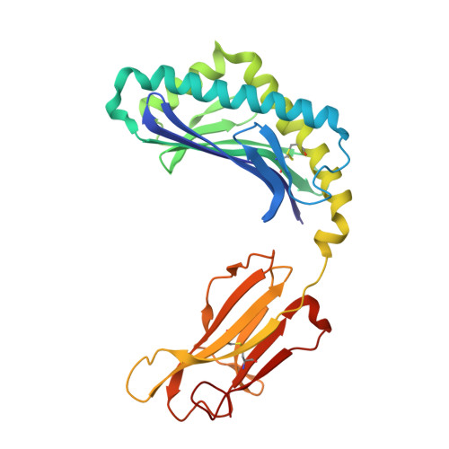
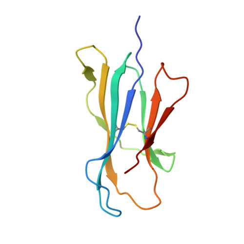
Cholesteryl esters stabilize human CD1c conformations for recognition by self-reactive T cells.

Mansour, S.Tocheva, A.S.Cave-Ayland, C.Machelett, M.M.Sander, B.Lissin, N.M.Molloy, P.E.Baird, M.S.Stubs, G.Schroder, N.W.Schumann, R.R.Rademann, J.Postle, A.D.Jakobsen, B.K.Marshall, B.G.Gosain, R.Elkington, P.T.Elliott, T.Skylaris, C.K.Essex, J.W.Tews, I.Gadola, S.D.

(2016) Proc Natl Acad Sci U S A 113: E1266-E1275

  • DOI: https://doi.org/10.1073/pnas.1519246113
  • Primary Citation of Related Structures:  
    5C9J

  • PubMed Abstract: 

    Cluster of differentiation 1c (CD1c)-dependent self-reactive T cells are abundant in human blood, but self-antigens presented by CD1c to the T-cell receptors of these cells are poorly understood. Here we present a crystal structure of CD1c determined at 2.4 Å revealing an extended ligand binding potential of the antigen groove and a substantially different conformation compared with known CD1c structures. Computational simulations exploring different occupancy states of the groove reenacted these different CD1c conformations and suggested cholesteryl esters (CE) and acylated steryl glycosides (ASG) as new ligand classes for CD1c. Confirming this, we show that binding of CE and ASG to CD1c enables the binding of human CD1c self-reactive T-cell receptors. Hence, human CD1c adopts different conformations dependent on ligand occupancy of its groove, with CE and ASG stabilizing CD1c conformations that provide a footprint for binding of CD1c self-reactive T-cell receptors.


  • Organizational Affiliation

    Clinical and Experimental Sciences, Faculty of Medicine, University of Southampton, Southampton SO17 1BJ, United Kingdom; Institute for Life Sciences, University of Southampton, Southampton SO17 1BJ, United Kingdom; sgadola@gmail.com s.mansour@soton.ac.uk.


Macromolecules
Find similar proteins by:  (by identity cutoff)  |  3D Structure
Entity ID: 1
MoleculeChains Sequence LengthOrganismDetailsImage
T-cell surface glycoprotein CD1c,T-cell surface glycoprotein CD1b281Homo sapiensMutation(s): 0 
Gene Names: CD1CCD1B
UniProt & NIH Common Fund Data Resources
Find proteins for P29016 (Homo sapiens)
Explore P29016 
Go to UniProtKB:  P29016
PHAROS:  P29016
GTEx:  ENSG00000158485 
Find proteins for P29017 (Homo sapiens)
Explore P29017 
Go to UniProtKB:  P29017
PHAROS:  P29017
GTEx:  ENSG00000158481 
Entity Groups  
Sequence Clusters30% Identity50% Identity70% Identity90% Identity95% Identity100% Identity
UniProt GroupsP29017P29016
Sequence Annotations
Expand
  • Reference Sequence
Find similar proteins by:  (by identity cutoff)  |  3D Structure
Entity ID: 2
MoleculeChains Sequence LengthOrganismDetailsImage
Beta-2-microglobulin99Homo sapiensMutation(s): 0 
Gene Names: B2MCDABP0092HDCMA22P
UniProt & NIH Common Fund Data Resources
Find proteins for P61769 (Homo sapiens)
Explore P61769 
Go to UniProtKB:  P61769
PHAROS:  P61769
GTEx:  ENSG00000166710 
Entity Groups  
Sequence Clusters30% Identity50% Identity70% Identity90% Identity95% Identity100% Identity
UniProt GroupP61769
Sequence Annotations
Expand
  • Reference Sequence
Small Molecules
Ligands 4 Unique
IDChains Name / Formula / InChI Key2D Diagram3D Interactions
STE
Query on STE

Download Ideal Coordinates CCD File 
E [auth A]STEARIC ACID
C18 H36 O2
QIQXTHQIDYTFRH-UHFFFAOYSA-N
DAO
Query on DAO

Download Ideal Coordinates CCD File 
C [auth A],
D [auth A]
LAURIC ACID
C12 H24 O2
POULHZVOKOAJMA-UHFFFAOYSA-N
PO4
Query on PO4

Download Ideal Coordinates CCD File 
I [auth A],
J [auth A]
PHOSPHATE ION
O4 P
NBIIXXVUZAFLBC-UHFFFAOYSA-K
GOL
Query on GOL

Download Ideal Coordinates CCD File 
F [auth A],
G [auth A],
H [auth A],
K [auth B]
GLYCEROL
C3 H8 O3
PEDCQBHIVMGVHV-UHFFFAOYSA-N
Experimental Data & Validation

Experimental Data

  • Method: X-RAY DIFFRACTION
  • Resolution: 2.40 Å
  • R-Value Free: 0.200 
  • R-Value Work: 0.155 
  • R-Value Observed: 0.158 
  • Space Group: P 31 2 1
Unit Cell:
Length ( Å )Angle ( ˚ )
a = 97.13α = 90
b = 97.13β = 90
c = 115.08γ = 120
Software Package:
Software NamePurpose
REFMACrefinement
Cootmodel building
PHASERphasing
XSCALEdata scaling
XDSdata reduction

Structure Validation

View Full Validation Report


Ligand Structure Quality Assessment 


Entry History & Funding Information

Deposition Data


Funding OrganizationLocationGrant Number
HEFCEUnited Kingdom--

Revision History  (Full details and data files)

  • Version 1.0: 2016-03-02
    Type: Initial release
  • Version 1.1: 2016-03-09
    Changes: Database references
  • Version 1.2: 2024-01-10
    Changes: Data collection, Database references, Refinement description