5B0V

Crystal Structure of Marburg virus VP40 Dimer


Experimental Data Snapshot

  • Method: X-RAY DIFFRACTION
  • Resolution: 2.81 Å
  • R-Value Free: 0.257 
  • R-Value Work: 0.202 
  • R-Value Observed: 0.205 

wwPDB Validation   3D Report Full Report


This is version 1.1 of the entry. See complete history


Literature

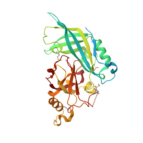
Crystal Structure of Marburg Virus VP40 Reveals a Broad, Basic Patch for Matrix Assembly and a Requirement of the N-Terminal Domain for Immunosuppression

Oda, S.Noda, T.Wijesinghe, K.J.Halfmann, P.Bornholdt, Z.A.Abelson, D.M.Armbrust, T.Stahelin, R.V.Kawaoka, Y.Saphire, E.O.

(2015) J Virol 90: 1839-1848

  • DOI: https://doi.org/10.1128/JVI.01597-15
  • Primary Citation of Related Structures:  
    5B0V

  • PubMed Abstract: 

    Marburg virus (MARV), a member of the filovirus family, causes severe hemorrhagic fever with up to 90% lethality. MARV matrix protein VP40 is essential for assembly and release of newly copied viruses and also suppresses immune signaling in the infected cell. Here we report the crystal structure of MARV VP40. We found that MARV VP40 forms a dimer in solution, mediated by N-terminal domains, and that formation of this dimer is essential for budding of virus-like particles. We also found the N-terminal domain to be necessary and sufficient for immune antagonism. The C-terminal domains of MARV VP40 are dispensable for immunosuppression but are required for virus assembly. The C-terminal domains are only 16% identical to those of Ebola virus, differ in structure from those of Ebola virus, and form a distinct broad and flat cationic surface that likely interacts with the cell membrane during virus assembly.


  • Organizational Affiliation

    Department of Immunology and Microbial Science, The Scripps Research Institute, La Jolla, California, USA.


Macromolecules
Find similar proteins by:  (by identity cutoff)  |  3D Structure
Entity ID: 1
MoleculeChains Sequence LengthOrganismDetailsImage
Matrix protein VP40
A, B
317Marburg virus - Musoke, Kenya, 1980Mutation(s): 0 
Gene Names: VP40
UniProt
Find proteins for P35260 (Lake Victoria marburgvirus (strain Musoke-80))
Explore P35260 
Go to UniProtKB:  P35260
Entity Groups  
Sequence Clusters30% Identity50% Identity70% Identity90% Identity95% Identity100% Identity
UniProt GroupP35260
Sequence Annotations
Expand
  • Reference Sequence
Small Molecules
Ligands 1 Unique
IDChains Name / Formula / InChI Key2D Diagram3D Interactions
EOH
Query on EOH

Download Ideal Coordinates CCD File 
C [auth B]ETHANOL
C2 H6 O
LFQSCWFLJHTTHZ-UHFFFAOYSA-N
Modified Residues  1 Unique
IDChains TypeFormula2D DiagramParent
MSE
Query on MSE
A, B
L-PEPTIDE LINKINGC5 H11 N O2 SeMET
Experimental Data & Validation

Experimental Data

  • Method: X-RAY DIFFRACTION
  • Resolution: 2.81 Å
  • R-Value Free: 0.257 
  • R-Value Work: 0.202 
  • R-Value Observed: 0.205 
  • Space Group: I 2 2 2
Unit Cell:
Length ( Å )Angle ( ˚ )
a = 95.81α = 90
b = 107.31β = 90
c = 127.65γ = 90
Software Package:
Software NamePurpose
REFMACrefinement
HKL-2000data reduction
HKL-2000data scaling
Auto-Rickshawphasing

Structure Validation

View Full Validation Report


Entry History 

Deposition Data

Revision History  (Full details and data files)

  • Version 1.0: 2016-01-20
    Type: Initial release
  • Version 1.1: 2016-03-09
    Changes: Database references