5AON

Crystal structure of the conserved N-terminal domain of Pex14 from Trypanosoma brucei


Experimental Data Snapshot

  • Method: X-RAY DIFFRACTION
  • Resolution: 1.65 Å
  • R-Value Free: 0.232 
  • R-Value Work: 0.195 
  • R-Value Observed: 0.197 

wwPDB Validation   3D Report Full Report


This is version 1.3 of the entry. See complete history


Literature

Characterization of the Interaction between Trypanosoma Brucei Pex5P and its Receptor Pex14P.

Watanabe, Y.Kawaguchi, K.Okuyama, N.Sugawara, Y.Obita, T.Mizuguchi, M.Morita, M.Imanaka, T.

(2016) FEBS Lett 590: 242

  • DOI: https://doi.org/10.1002/1873-3468.12044
  • Primary Citation of Related Structures:  
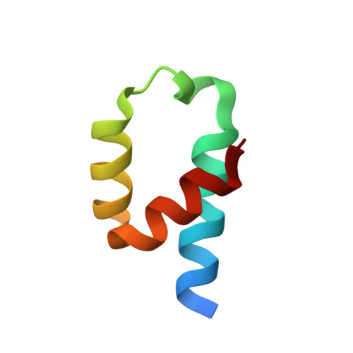
    5AON

  • PubMed Abstract: 

    The interaction of Trypanosoma brucei (Tb) Pex5p and its receptor TbPex14p is essential for the translocation of newly synthesized matrix proteins into the glycosome. Here, we reveal that only the third WXXXF/Y motif of TbPex5p is involved in the interaction and that negative charge of the fourth amino acid is important. We suggest that Phe35 and Phe52 of TbPex14p interact with Trp318 and Phe322 in the third motif and that the Lys56 adjacent to Phe35/Phe52 associates with the fourth Glu in the motif to make the complex. This information is expected to be useful for developing anti-trypanosomal drugs.


  • Organizational Affiliation

    Department of Biological Chemistry, Graduate School of Medicine and Pharmaceutical Sciences, University of Toyama, Sugitani, Japan.


Macromolecules
Find similar proteins by:  (by identity cutoff)  |  3D Structure
Entity ID: 1
MoleculeChains Sequence LengthOrganismDetailsImage
PEROXIN 14
A, B, C, D, E
53Trypanosoma bruceiMutation(s): 0 
UniProt
Find proteins for Q8IEW2 (Trypanosoma brucei brucei)
Explore Q8IEW2 
Go to UniProtKB:  Q8IEW2
Entity Groups  
Sequence Clusters30% Identity50% Identity70% Identity90% Identity95% Identity100% Identity
UniProt GroupQ8IEW2
Sequence Annotations
Expand
  • Reference Sequence
Experimental Data & Validation

Experimental Data

  • Method: X-RAY DIFFRACTION
  • Resolution: 1.65 Å
  • R-Value Free: 0.232 
  • R-Value Work: 0.195 
  • R-Value Observed: 0.197 
  • Space Group: P 61 2 2
Unit Cell:
Length ( Å )Angle ( ˚ )
a = 53.569α = 90
b = 53.569β = 90
c = 342.827γ = 120
Software Package:
Software NamePurpose
PHENIXrefinement
MOSFLMdata reduction
SCALAdata scaling
PHASERphasing

Structure Validation

View Full Validation Report


Entry History 

Deposition Data

Revision History  (Full details and data files)

  • Version 1.0: 2015-12-23
    Type: Initial release
  • Version 1.1: 2016-01-27
    Changes: Database references
  • Version 1.2: 2016-02-10
    Changes: Database references
  • Version 1.3: 2024-01-10
    Changes: Data collection, Database references, Derived calculations, Other, Refinement description