Structural Adaptations of Norovirus GII.17/13/21 Lineage through Two Distinct Evolutionary Paths.
Qian, Y., Song, M., Jiang, X., Xia, M., Meller, J., Tan, M., Chen, Y., Li, X., Rao, Z.(2019) J Virol 93
- PubMed: 30333166
- DOI: https://doi.org/10.1128/JVI.01655-18
- Primary Citation of Related Structures:
5ZUQ, 5ZUS, 5ZV9 - PubMed Abstract:
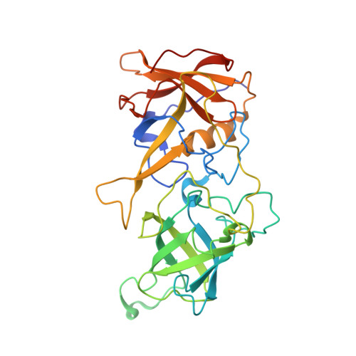
Human noroviruses (huNoVs), which cause epidemic acute gastroenteritis, recognize histo-blood group antigens (HBGAs) as host attachment factors affecting host susceptibility. HuNoVs are genetically diverse, containing at least 31 genotypes in the two major genogroups (genogroup I [GI] and GII). Three GII genotypes, GII genotype 17 (GII.17), GII.13, and GII.21, form a unique genetic lineage, in which the GII.17 genotype retains the conventional GII HBGA binding site (HBS), while the GII.13/21 genotypes acquire a completely new HBS. To understand the molecular bases behind these evolutionary changes, we solved the crystal structures of the HBGA binding protruding domains of (i) an early GII.17 variant (the 1978 variant) that does not bind or binds weakly to HBGAs, (ii) the new GII.17 variant (the 2014/15 variant) that binds A/B/H antigens strongly via an optimized GII HBS, and (iii) a GII.13 variant (the 2010 variant) that binds the Lewis a (Le a ) antigen via the new HBS. These serial, high-resolution structural data enable a comprehensive structural comparison to understand the evolutionary changes of the GII.17/13/21 lineage, including the emergence of the new HBS of the GII.13/21 sublineage and the possible HBS optimization of the recent GII.17 variant for an enhanced HBGA binding ability. Our study elucidates the structural adaptations of the GII.17/13/21 lineage through distinct evolutionary paths, which may allow a theory explaining huNoV adaptations and evolutions to be put forward. IMPORTANCE Our understanding of the molecular bases behind the interplays between human noroviruses and their host glycan ligands, as well as their evolutionary changes over time with alterations in their host ligand binding capability and host susceptibility, remains limited. By solving the crystal structures of the glycan ligand binding protruding (P) domains with or without glycan ligands of three representative noroviruses of the GII.17/13/21 genetic lineage, we elucidated the molecular bases of the human norovirus-glycan interactions of this special genetic lineage. We present solid evidence on how noroviruses of this genetic lineage evolved via different evolutionary paths to (i) optimize their glycan binding site for higher glycan binding function and (ii) acquire a completely new glycan binding site for new ligands. Our data shed light on the mechanism of the structural adaptations of human noroviruses through different evolutionary paths, facilitating our understanding of human norovirus adaptations, evolutions, and epidemiology.
Organizational Affiliation:
National Laboratory of Biomacromolecules, Institute of Biophysics, Chinese Academy of Sciences, Beijing, China.


