5ZUR

Achromobacter Dh1f Bacterioferritin


Experimental Data Snapshot

  • Method: X-RAY DIFFRACTION
  • Resolution: 1.95 Å
  • R-Value Free: 0.187 
  • R-Value Work: 0.142 
  • R-Value Observed: 0.144 

wwPDB Validation   3D Report Full Report


Ligand Structure Quality Assessment 


This is version 1.2 of the entry. See complete history


Literature

Serendipitous crystallization and structure determination of bacterioferritin from Achromobacter.

Dwivedy, A.Jha, B.Singh, K.H.Ahmad, M.Ashraf, A.Kumar, D.Biswal, B.K.

(2018) Acta Crystallogr F Struct Biol Commun 74: 558-566

  • DOI: https://doi.org/10.1107/S2053230X18009809
  • Primary Citation of Related Structures:  
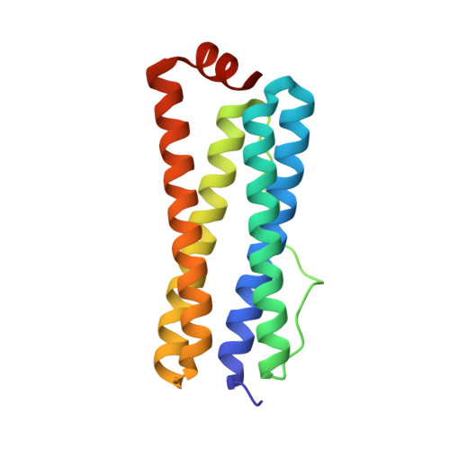
    5ZUR

  • PubMed Abstract: 

    Bacterioferritins (Bfrs) are ferritin-like molecules with a hollow spherical 24-mer complex design that are unique to bacterial and archaeal species. They play a critical role in storing iron(III) within the complex at concentrations much higher than the feasible solubility limits of iron(III), thus maintaining iron homeostasis within cells. Here, the crystal structure of bacterioferritin from Achromobacter (Ach Bfr) that crystallized serendipitously during a crystallization attempt of an unrelated mycobacterial protein is reported at 1.95 Å resolution. Notably, Fe atoms were bound to the structure along with a porphyrin ring sandwiched between the subunits of a dimer. Furthermore, the dinuclear ferroxidase center of Ach Bfr has only a single iron bound, in contrast to the two Fe atoms in other Bfrs. The structure of Ach Bfr clearly demonstrates the substitution of a glutamate residue, which is involved in the interaction with the second Fe atom, by a threonine and the consequent absence of another Fe atom there. The iron at the dinuclear center has a tetravalent coordination, while a second iron with a hexavalent coordination was found within the porphyrin ring, generating a heme moiety. Achromobacter spp. are known opportunistic pathogens; this structure enhances the current understanding of their iron metabolism and regulation, and importantly will be useful in the design of small-molecule inhibitors against this protein through a structure-guided approach.


  • Organizational Affiliation

    Structural and Functional Biology Laboratory, National Institute of Immunology, Aruna Asaf Ali Marg, New Delhi 110 067, India.


Macromolecules
Find similar proteins by:  (by identity cutoff)  |  3D Structure
Entity ID: 1
MoleculeChains Sequence LengthOrganismDetailsImage
Bacterioferritin
A, B, C
158Achromobacter sp. DH1fMutation(s): 0 
UniProt
Find proteins for A0A3F2YLV4 (Achromobacter sp. DH1f)
Explore A0A3F2YLV4 
Go to UniProtKB:  A0A3F2YLV4
Entity Groups  
Sequence Clusters30% Identity50% Identity70% Identity90% Identity95% Identity100% Identity
UniProt GroupA0A3F2YLV4
Sequence Annotations
Expand
  • Reference Sequence
Small Molecules
Ligands 5 Unique
IDChains Name / Formula / InChI Key2D Diagram3D Interactions
HEM
Query on HEM

Download Ideal Coordinates CCD File 
D [auth A],
K [auth B],
P [auth C]
PROTOPORPHYRIN IX CONTAINING FE
C34 H32 Fe N4 O4
KABFMIBPWCXCRK-RGGAHWMASA-L
BA
Query on BA

Download Ideal Coordinates CCD File 
H [auth A],
M [auth B],
R [auth C]
BARIUM ION
Ba
XDFCIPNJCBUZJN-UHFFFAOYSA-N
PEG
Query on PEG

Download Ideal Coordinates CCD File 
F [auth A],
G [auth A],
L [auth B],
O [auth C]
DI(HYDROXYETHYL)ETHER
C4 H10 O3
MTHSVFCYNBDYFN-UHFFFAOYSA-N
FE2
Query on FE2

Download Ideal Coordinates CCD File 
E [auth A],
J [auth B],
Q [auth C]
FE (II) ION
Fe
CWYNVVGOOAEACU-UHFFFAOYSA-N
CL
Query on CL

Download Ideal Coordinates CCD File 
I [auth A],
N [auth B],
S [auth C]
CHLORIDE ION
Cl
VEXZGXHMUGYJMC-UHFFFAOYSA-M
Experimental Data & Validation

Experimental Data

  • Method: X-RAY DIFFRACTION
  • Resolution: 1.95 Å
  • R-Value Free: 0.187 
  • R-Value Work: 0.142 
  • R-Value Observed: 0.144 
  • Space Group: I 4 2 2
Unit Cell:
Length ( Å )Angle ( ˚ )
a = 158.665α = 90
b = 158.665β = 90
c = 118.061γ = 90
Software Package:
Software NamePurpose
REFMACrefinement
HKL-2000data reduction
HKL-2000data scaling
PHASERphasing

Structure Validation

View Full Validation Report


Ligand Structure Quality Assessment 


Entry History 

Deposition Data

Revision History  (Full details and data files)

  • Version 1.0: 2018-09-05
    Type: Initial release
  • Version 1.1: 2018-09-19
    Changes: Data collection, Database references
  • Version 1.2: 2023-11-22
    Changes: Data collection, Database references, Derived calculations, Refinement description