5ZRR

Crystal structure of PET-degrading cutinase Cut190 S176A/S226P/R228S mutant in monoethyl succinate bound state


Experimental Data Snapshot

  • Method: X-RAY DIFFRACTION
  • Resolution: 1.34 Å
  • R-Value Free: 0.185 
  • R-Value Work: 0.162 
  • R-Value Observed: 0.164 

wwPDB Validation   3D Report Full Report


Ligand Structure Quality Assessment 


This is version 1.2 of the entry. See complete history


Literature

Structural Dynamics of the PET-Degrading Cutinase-like Enzyme from Saccharomonospora viridis AHK190 in Substrate-Bound States Elucidates the Ca2+-Driven Catalytic Cycle.

Numoto, N.Kamiya, N.Bekker, G.J.Yamagami, Y.Inaba, S.Ishii, K.Uchiyama, S.Kawai, F.Ito, N.Oda, M.

(2018) Biochemistry 57: 5289-5300

  • DOI: https://doi.org/10.1021/acs.biochem.8b00624
  • Primary Citation of Related Structures:  
    5ZNO, 5ZRQ, 5ZRR, 5ZRS

  • PubMed Abstract: 

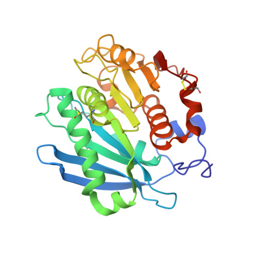
    A cutinase-type polyesterase from Saccharomonospora viridis AHK190 (Cut190) has been shown to degrade the inner block of polyethylene terephthalate. A unique feature of Cut190 is that its function and stability are regulated by Ca 2+ binding. Our previous crystal structure analysis of Cut190S226P showed that one Ca 2+ binds to the enzyme, which induces large conformational changes in several loop regions to stabilize an open conformation [Miyakawa, T., et al. (2015) Appl. Microbiol. Biotechnol. 99, 4297]. In this study, to analyze the substrate recognition mechanism of Cut190, we determined the crystal structure of the inactive form of a Cut190 mutant, Cut190*S176A, in complex with calcium ions and/or substrates. We found that three calcium ions bind to Cut190*S176A, which is supported by analysis using native mass spectrometry experiments and 3D Reference Interaction Site Model calculations. The complex structures with the two substrates, monoethyl succinate and monoethyl adipate (engaged and open forms), presumably correspond to the pre- and post-reaction states, as the ester bond is close to the active site and pointing outward from the active site, respectively, for the two complexes. Ca 2+ binding induces the pocket to open, enabling the substrate to access the pocket more easily. Molecular dynamics simulations suggest that a post-reaction state in the engaged form presumably exists between the experimentally observed forms, indicating that the substrate would be cleaved in the engaged form and then requires the enzyme to change to the open form to release the product, a process that Ca 2+ can greatly accelerate.


  • Organizational Affiliation

    Medical Research Institute , Tokyo Medical and Dental University , 1-5-45 Yushima , Bunkyo-ku, Tokyo 113-8510 , Japan.


Macromolecules
Find similar proteins by:  (by identity cutoff)  |  3D Structure
Entity ID: 1
MoleculeChains Sequence LengthOrganismDetailsImage
Alpha/beta hydrolase family protein265Saccharomonospora viridisMutation(s): 3 
Gene Names: Cut190SAMN02982918_2340
UniProt
Find proteins for W0TJ64 (Saccharomonospora viridis)
Explore W0TJ64 
Go to UniProtKB:  W0TJ64
Entity Groups  
Sequence Clusters30% Identity50% Identity70% Identity90% Identity95% Identity100% Identity
UniProt GroupW0TJ64
Sequence Annotations
Expand
  • Reference Sequence
Small Molecules
Ligands 4 Unique
IDChains Name / Formula / InChI Key2D Diagram3D Interactions
9J3 (Subject of Investigation/LOI)
Query on 9J3

Download Ideal Coordinates CCD File 
B [auth A]4-ethoxy-4-oxobutanoic acid
C6 H10 O4
LOLKAJARZKDJTD-UHFFFAOYSA-N
SO4
Query on SO4

Download Ideal Coordinates CCD File 
H [auth A]SULFATE ION
O4 S
QAOWNCQODCNURD-UHFFFAOYSA-L
GOL
Query on GOL

Download Ideal Coordinates CCD File 
G [auth A]GLYCEROL
C3 H8 O3
PEDCQBHIVMGVHV-UHFFFAOYSA-N
ZN
Query on ZN

Download Ideal Coordinates CCD File 
C [auth A],
D [auth A],
E [auth A],
F [auth A]
ZINC ION
Zn
PTFCDOFLOPIGGS-UHFFFAOYSA-N
Modified Residues  1 Unique
IDChains TypeFormula2D DiagramParent
MHO
Query on MHO
A
L-PEPTIDE LINKINGC5 H11 N O3 SMET
Experimental Data & Validation

Experimental Data

  • Method: X-RAY DIFFRACTION
  • Resolution: 1.34 Å
  • R-Value Free: 0.185 
  • R-Value Work: 0.162 
  • R-Value Observed: 0.164 
  • Space Group: P 21 21 21
Unit Cell:
Length ( Å )Angle ( ˚ )
a = 49.507α = 90
b = 61.397β = 90
c = 76.783γ = 90
Software Package:
Software NamePurpose
PHENIXrefinement
XDSdata reduction
XDSdata scaling
PHASERphasing

Structure Validation

View Full Validation Report


Ligand Structure Quality Assessment 


Entry History & Funding Information

Deposition Data


Funding OrganizationLocationGrant Number
Japan Agency for Medical Research and Development (AMED)JapanJP17am0101001

Revision History  (Full details and data files)

  • Version 1.0: 2018-09-12
    Type: Initial release
  • Version 1.1: 2018-09-26
    Changes: Data collection, Database references
  • Version 1.2: 2023-11-22
    Changes: Data collection, Database references, Derived calculations, Refinement description