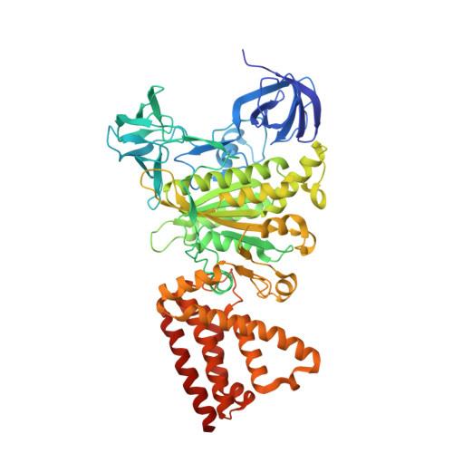
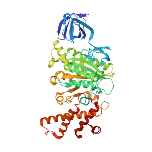
5ZE9

Crystal structure of AMP-PNP bound mutant A3B3 complex from Enterococcus hirae V-ATPase


Experimental Data Snapshot

  • Method: X-RAY DIFFRACTION
  • Resolution: 2.10 Å
  • R-Value Free: 0.196 
  • R-Value Work: 0.166 
  • R-Value Observed: 0.168 

wwPDB Validation   3D Report Full Report


Ligand Structure Quality Assessment 


This is version 1.2 of the entry. See complete history


Literature

Metastable asymmetrical structure of a shaftless V1motor.

Maruyama, S.Suzuki, K.Imamura, M.Sasaki, H.Matsunami, H.Mizutani, K.Saito, Y.Imai, F.L.Ishizuka-Katsura, Y.Kimura-Someya, T.Shirouzu, M.Uchihashi, T.Ando, T.Yamato, I.Murata, T.

(2019) Sci Adv 5: eaau8149-eaau8149

  • DOI: https://doi.org/10.1126/sciadv.aau8149
  • Primary Citation of Related Structures:  
    5ZE9, 5ZEA

  • PubMed Abstract: 

    V 1 -ATPase is an ATP-driven rotary motor that is composed of a ring-shaped A 3 B 3 complex and a central DF shaft. The nucleotide-free A 3 B 3 complex of Enterococcus hirae , composed of three identical A 1 B 1 heterodimers, showed a unique asymmetrical structure, probably due to the strong binding of the N-terminal barrel domain, which forms a crown structure. Here, we mutated the barrel region to weaken the crown, and performed structural analyses using high-speed atomic force microscopy and x-ray crystallography of the mutant A 3 B 3 . The nucleotide-free mutant A 3 B 3 complex had a more symmetrical open structure than the wild type. Binding of nucleotides produced a closely packed spiral-like structure with a disrupted crown. These findings suggest that wild-type A 3 B 3 forms a metastable (stressed) asymmetric structure composed of unstable A 1 B 1 conformers due to the strong constraint of the crown. The results further the understanding of the principle of the cooperative transition mechanism of rotary motors.


  • Organizational Affiliation

    Department of Chemistry, Graduate School of Science, Chiba University, 1-33 Yayoi-cho, Inage, Chiba 263-8522, Japan.


Macromolecules
Find similar proteins by:  (by identity cutoff)  |  3D Structure
Entity ID: 1
MoleculeChains Sequence LengthOrganismDetailsImage
V-type sodium ATPase catalytic subunit A
A, B, C
600Enterococcus hirae ATCC 9790Mutation(s): 0 
Gene Names: ntpAEHR_08260
EC: 3.6.3.15
UniProt
Find proteins for Q08636 (Enterococcus hirae (strain ATCC 9790 / DSM 20160 / JCM 8729 / LMG 6399 / NBRC 3181 / NCIMB 6459 / NCDO 1258 / NCTC 12367 / WDCM 00089 / R))
Explore Q08636 
Go to UniProtKB:  Q08636
Entity Groups  
Sequence Clusters30% Identity50% Identity70% Identity90% Identity95% Identity100% Identity
UniProt GroupQ08636
Sequence Annotations
Expand
  • Reference Sequence
Find similar proteins by:  (by identity cutoff)  |  3D Structure
Entity ID: 2
MoleculeChains Sequence LengthOrganismDetailsImage
V-type sodium ATPase subunit B
D, E, F
465Enterococcus hirae ATCC 9790Mutation(s): 1 
Gene Names: ntpBEHR_08265
UniProt
Find proteins for Q08637 (Enterococcus hirae (strain ATCC 9790 / DSM 20160 / JCM 8729 / LMG 6399 / NBRC 3181 / NCIMB 6459 / NCDO 1258 / NCTC 12367 / WDCM 00089 / R))
Explore Q08637 
Go to UniProtKB:  Q08637
Entity Groups  
Sequence Clusters30% Identity50% Identity70% Identity90% Identity95% Identity100% Identity
UniProt GroupQ08637
Sequence Annotations
Expand
  • Reference Sequence
Small Molecules
Ligands 4 Unique
IDChains Name / Formula / InChI Key2D Diagram3D Interactions
ANP
Query on ANP

Download Ideal Coordinates CCD File 
BA [auth C],
G [auth A],
P [auth B]
PHOSPHOAMINOPHOSPHONIC ACID-ADENYLATE ESTER
C10 H17 N6 O12 P3
PVKSNHVPLWYQGJ-KQYNXXCUSA-N
MES
Query on MES

Download Ideal Coordinates CCD File 
LA [auth D]2-(N-MORPHOLINO)-ETHANESULFONIC ACID
C6 H13 N O4 S
SXGZJKUKBWWHRA-UHFFFAOYSA-N
GOL
Query on GOL

Download Ideal Coordinates CCD File 
AA [auth B]
DA [auth C]
EA [auth C]
FA [auth C]
GA [auth C]
AA [auth B],
DA [auth C],
EA [auth C],
FA [auth C],
GA [auth C],
HA [auth C],
I [auth A],
IA [auth C],
J [auth A],
JA [auth C],
K [auth A],
KA [auth C],
L [auth A],
M [auth A],
MA [auth D],
N [auth A],
NA [auth D],
O [auth A],
OA [auth D],
PA [auth E],
QA [auth E],
R [auth B],
RA [auth E],
S [auth B],
SA [auth E],
T [auth B],
TA [auth F],
U [auth B],
UA [auth F],
V [auth B],
VA [auth F],
W [auth B],
WA [auth F],
X [auth B],
XA [auth F],
Y [auth B],
YA [auth F],
Z [auth B],
ZA [auth F]
GLYCEROL
C3 H8 O3
PEDCQBHIVMGVHV-UHFFFAOYSA-N
MG
Query on MG

Download Ideal Coordinates CCD File 
CA [auth C],
H [auth A],
Q [auth B]
MAGNESIUM ION
Mg
JLVVSXFLKOJNIY-UHFFFAOYSA-N
Experimental Data & Validation

Experimental Data

  • Method: X-RAY DIFFRACTION
  • Resolution: 2.10 Å
  • R-Value Free: 0.196 
  • R-Value Work: 0.166 
  • R-Value Observed: 0.168 
  • Space Group: P 21 21 21
Unit Cell:
Length ( Å )Angle ( ˚ )
a = 105.83α = 90
b = 151.37β = 90
c = 235.49γ = 90
Software Package:
Software NamePurpose
XDSdata reduction
HKL-2000data scaling
MOLREPphasing
PHENIXrefinement
PDB_EXTRACTdata extraction

Structure Validation

View Full Validation Report


Ligand Structure Quality Assessment 


Entry History 

Deposition Data

Revision History  (Full details and data files)

  • Version 1.0: 2019-02-06
    Type: Initial release
  • Version 1.1: 2019-02-20
    Changes: Data collection, Database references
  • Version 1.2: 2023-11-22
    Changes: Data collection, Database references, Derived calculations, Refinement description