5Z2F

NADPH/PDA bound Dihydrodipicolinate reductase from Paenisporosarcina sp. TG-14


Experimental Data Snapshot

  • Method: X-RAY DIFFRACTION
  • Resolution: 2.10 Å
  • R-Value Free: 0.257 
  • R-Value Work: 0.187 
  • R-Value Observed: 0.190 

wwPDB Validation   3D Report Full Report


Ligand Structure Quality Assessment 


This is version 1.1 of the entry. See complete history


Literature

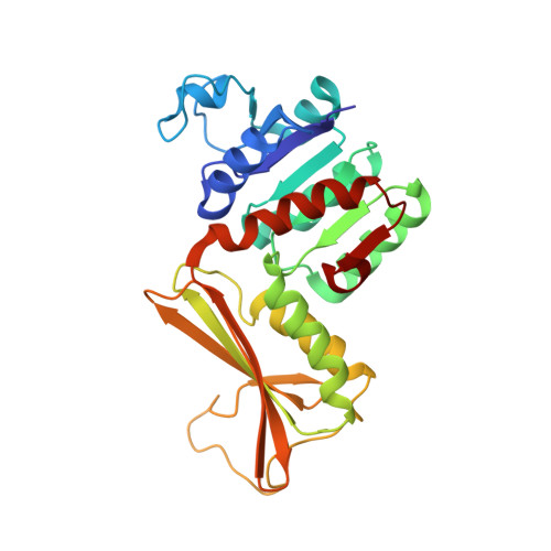
Crystal structure of dihydrodipicolinate reductase (PaDHDPR) from Paenisporosarcina sp. TG-14: structural basis for NADPH preference as a cofactor

Lee, C.W.Park, S.H.Lee, S.G.Park, H.H.Kim, H.J.Park, H.Park, H.Lee, J.H.

(2018) Sci Rep 8: 7936-7936

  • DOI: https://doi.org/10.1038/s41598-018-26291-x
  • Primary Citation of Related Structures:  
    5Z2D, 5Z2E, 5Z2F

  • PubMed Abstract: 

    Dihydrodipicolinate reductase (DHDPR) is a key enzyme in the diaminopimelate- and lysine-synthesis pathways that reduces DHDP to tetrahydrodipicolinate. Although DHDPR uses both NADPH and NADH as a cofactor, the structural basis for cofactor specificity and preference remains unclear. Here, we report that Paenisporosarcina sp. TG-14 PaDHDPR has a strong preference for NADPH over NADH, as determined by isothermal titration calorimetry and enzymatic activity assays. We determined the crystal structures of PaDHDPR alone, with its competitive inhibitor (dipicolinate), and the ternary complex of the enzyme with dipicolinate and NADPH, with results showing that only the ternary complex had a fully closed conformation and suggesting that binding of both substrate and nucleotide cofactor is required for enzymatic activity. Moreover, NADPH binding induced local conformational changes in the N-terminal long loop (residues 34-59) of PaDHDPR, as the His35 and Lys36 residues in this loop interacted with the 2'-phosphate group of NADPH, possibly accounting for the strong preference of PaDHDPR for NADPH. Mutation of these residues revealed reduced NADPH binding and enzymatic activity, confirming their importance in NADPH binding. These findings provide insight into the mechanism of action and cofactor selectivity of this important bacterial enzyme.


  • Organizational Affiliation

    Unit of Polar Genomics, Korea Polar Research Institute, Incheon, 21990, Republic of Korea.


Macromolecules
Find similar proteins by:  (by identity cutoff)  |  3D Structure
Entity ID: 1
MoleculeChains Sequence LengthOrganismDetailsImage
Dihydrodipicolinate reductase265Paenisporosarcina sp. TG-14Mutation(s): 0 
UniProt
Find proteins for A0A3B6UER2 (Paenisporosarcina sp. TG-14)
Explore A0A3B6UER2 
Go to UniProtKB:  A0A3B6UER2
Entity Groups  
Sequence Clusters30% Identity50% Identity70% Identity90% Identity95% Identity100% Identity
UniProt GroupA0A3B6UER2
Sequence Annotations
Expand
  • Reference Sequence
Small Molecules
Ligands 2 Unique
IDChains Name / Formula / InChI Key2D Diagram3D Interactions
NAP
Query on NAP

Download Ideal Coordinates CCD File 
B [auth A]NADP NICOTINAMIDE-ADENINE-DINUCLEOTIDE PHOSPHATE
C21 H28 N7 O17 P3
XJLXINKUBYWONI-NNYOXOHSSA-N
PDC
Query on PDC

Download Ideal Coordinates CCD File 
C [auth A]PYRIDINE-2,6-DICARBOXYLIC ACID
C7 H5 N O4
WJJMNDUMQPNECX-UHFFFAOYSA-N
Experimental Data & Validation

Experimental Data

  • Method: X-RAY DIFFRACTION
  • Resolution: 2.10 Å
  • R-Value Free: 0.257 
  • R-Value Work: 0.187 
  • R-Value Observed: 0.190 
  • Space Group: P 42 21 2
Unit Cell:
Length ( Å )Angle ( ˚ )
a = 79.942α = 90
b = 79.942β = 90
c = 83.835γ = 90
Software Package:
Software NamePurpose
REFMACrefinement
HKL-2000data reduction
HKL-2000data scaling

Structure Validation

View Full Validation Report


Ligand Structure Quality Assessment 


Entry History 

Deposition Data

Revision History  (Full details and data files)

  • Version 1.0: 2018-06-27
    Type: Initial release
  • Version 1.1: 2024-03-27
    Changes: Data collection, Database references